Atrial myxoma
BASIC INFORMATION
DEFINITION
Atrial myxoma is the most common primary tumor of the heart. Impaired filling of the left ventricle may also be caused by a tumor in the left atrium. Atrial myxomas account for approximately 75% of these tumors in adults. Atrial myxomas are pedunculated tumors that can mimic mitral stenosis by prolapsing into the mitral valve (Fig. 16). Complaints (dyspnea, palpitations, cyanosis, syncope) that change with body position, appearing in certain positions and disappearing in others, are virtually pathognomonic for this tumor.
EPIDEMIOLOGY & DEMOGRAPHICS
• Atrial myxomas account for 50% of all primary tumors of the heart.
• Approximately 75% of myxomas arise from the left atrium in close relationship to the fossa ovalis. Up to 15% of myxomas arise from the right atrial, with the remaining 10% arising from the left ventricle or from multiple sites.
• Approximately 90% of atrial myxomas are sporadic.
• 10% of atrial myxomas have an autosomal dominant transmission called Carney’s syndrome.
• The prevalence of atrial myxoma is approximately 75 cases/1 million autopsies.
• Average age of sporadic cases is 56 yr.
• Average age of familial cases is 25 yr.
• 70% of sporadic cases occur in females.
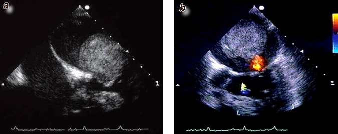
Fig. 16 Atrial myxoma
a) Transesophageal echocardiogram of a 68-year-old woman with a left-sided atrial myxoma; 2-D image in diastole. The left atrium is above, the left ventricle is below. The spherical myxoma enters the mitral valve during diastole, obstructing blood flow through the valve.
b) Color-coded Doppler echocardiogram of the same patient in systole. The myxoma is back in the left atrium. There is minimal “flame-shaped” backflow through the otherwise closed mitral valve.
PHYSICAL FINDINGS & CLINICAL PRESENTATION
Clinical Features. Besides a diastolic murmur, auscultation reveals an extra systolic sound or tumor plop caused by prolapse of the myxoma during systole. Changing the patient’s position alters the intensity of the audible findings. This does not occur in mitral stenosis. The diagnosis is established by echocardiography, which can positively distinguish atrial myxoma from mitral stenosis. Atrial myxoma is associated with noncardiac systemic symptoms such as fever (50 %), weight loss (25 %), dizziness and presyncope (20 %), anemia, signs of inflammation, and splenomegaly.
Clinical Features. Besides a diastolic murmur, auscultation reveals an extra systolic sound or tumor plop caused by prolapse of the myxoma during systole. Changing the patient’s position alters the intensity of the audible findings. This does not occur in mitral stenosis. The diagnosis is established by echocardiography, which can positively distinguish atrial myxoma from mitral stenosis. Atrial myxoma is associated with noncardiac systemic symptoms such as fever (50 %), weight loss (25 %), dizziness and presyncope (20 %), anemia, signs of inflammation, and splenomegaly.
Chronic signs of inflammation result from the production of inflammatory cytokines by the tumor itself. Atrial myxomas may lead to the systemic embolization of tumor fragments or thrombi. These emboli, which are usually multiple, may mimic vasculitis or infectious endocarditis.
Patients with atrial myxomas characteristically present in one of three ways:
• Mechanical valve obstruction (e.g., mitral or tricuspid valve)
1. Dyspnea on exertion
2. Orthopnea
3. Paroxysmal nocturnal dyspnea
4. Edema
5. Dizziness, lightheadedness, or syncope
6. Elevated jugular venous pressure
7. Loud S1, increased intensity of the P2 component of S2 secondary to pulmonary hypertension
8. Systolic murmurs of mitral regurgitation or tricuspid regurgitation and diastolic murmurs or mitral stenosis or tricuspid stenosis, depending on which chamber the myxoma arises from
9. Third heart sound called a “tumor plop”
10. Atrial fibrillation with an irregularly irregular pulse
• Systemic embolization may occur in up to 30% of cases leading to:
1. Cerebrovascular accidents
2. Pulmonary embolism
3. Paradoxical embolism
• Constitutional symptoms
1. Fever
2. Weight loss
3. Arthralgias
4. Raynaud phenomenon
ETIOLOGY
• Most cases (90%) of atrial myxomas are sporadic with no known cause. In the remaining 10% of cases a familial pattern occurs having an autosomal dominant transmission.
DIAGNOSIS
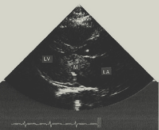
The diagnosis is easily made by echocardiography because the tumour is demonstrated as a dense spaceoccupying lesion (Fig. 16.1). Surgical removal usually results in a complete cure.
DIFFERENTIAL DIAGNOSIS
• Mitral stenosis
• Mitral regurgitaiton
• Tricuspid stenosis
• Tricuspid regurgitation
• Pulmonary hypertension
• Endocarditis
• Vasculitis
• Left atrial thrombus
• Pulmonary embolism
• Cerebrovascular accidents
• Collagen-vascular disease
• Carcinoid heart disease
• Ebstein’s anomaly
WORKUP
A high index of suspicion is needed to make the diagnosis of atrial myxoma as the clinical manifestations are similar to many common cardiovascular and pulmonary diseases.
LABORATORY TESTS
Although not very specific, the following laboratory values may be abnormal in patients with atrial myxomas:
• CBC: anemia, polycythemia, thrombocytopenia may occur
• Erythrocyte sedimentation rate, Creactive protein, and serum immunoglobulins are commonly elevated
• EKG: patients with atrial myxomas may have findings of left atrial enlargement, right atrial enlargement, atrial fibrillation, atrial flutter, premature ventricular
contractions, ventricular tachycardia, and ventricular fibrillation
Fig. 16.1 Atrial myxoma shown by a 2-D echocardiogram (long-axis view). The myxoma is an echo-dens mass obstructing the mitral valve orifice. It was removed surgically. LV, left ventricle; LA, left atrium; M, mass.
IMAGING STUDIES
• Chest x-ray examination: altered cardiac contour and chamber enlargement
• Echocardiography: initial imaging procedure of choice in suspected cases of atrial myxoma
• Transesophageal echocardiography: may pick up masses not visualized by transthoracic echocardiography
• MRI: Aids in delineating size, shape, and tumor characterizations
• Cardiac catheterization: usually not needed in diagnosing atrial myxoma; however, may be required in some circumstances to rule out other cardiac lesions (e.g., coronary artery disease)
TREATMENT
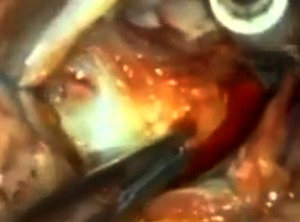
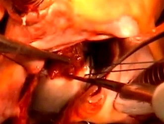
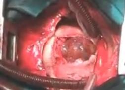
Cardiac Surgery (Fig. 16.2, 16.3, and 16.4)
NONPHARMACOLOGIC THERAPY
None
ACUTE GENERAL THERAPY
• Surgical excision is the treatment of choice.
• Surgery should be done promptly because sudden death can occur while waiting for the procedure (see “Disposition”).
CHRONIC Rx
Postoperative arrhythmias and conductions abnormalities were present in 26% of patients and can be treated according to standard convention.
DISPOSITION
• Surgical results have reported a 95% survival rate after a follow-up of 3 yr
• Up to 5% of sporadic cases of atrial myxoma may recur within the first 6 yr after surgery
• Up to 20% of familial cases of atrial myxoma may recur after surgery
• Sudden death has been reported to occur in up to 15% of patients with atrial myxoma, with death resulting from coronary or systemic embolization or by obstruction of blood flow at the mitral or tricuspid valve.
Fig. 16.2 Left Atrial Myxoma
Fig. 16.3 Left atrial myxoma-Cardiac Surgery
Fig. 16.4 Right Atrial Myxoma
Contacts: lubopitno_bg@abv.bg www.encyclopedia.lubopitko-bg.com Corporation. All rights reserved.
DON'T FORGET - KNOWLEDGE IS EVERYTHING!