Cellular Organization
Every human cell has a plasma membrane, a nucleus, and cytoplasm. The plasma membrane, which surrounds the cell and keeps it intact, regulates what enters and exits a cell. The plasma membrane is a phospholipid bilayer that is said to be semipermeable because it allows certain molecules but not others to enter the cell. Proteins present in the plasma membrane play important roles in allowing substances to enter the cell. The nucleus is a large, centrally located structure that can often be seen with a light microscope. The nucleus contains the chromosomes and is the control center of the cell. It controls the metabolic functioning and structural characteristics of the cell. The nucleolus is a region inside the nucleus.

The cytoplasm is the portion of the cell between the nucleus and the plasma membrane. The matrix of the cytoplasm is a semifluid medium that contains water and various types of molecules suspended or dissolved in the medium. The presence of proteins accounts for the semifluid nature of the matrix. The cytoplasm contains various organelles (Table 3.1 and Fig. 3.1).
Organelles are small, usually membranous structures that are best seen with an electron microscope. Each type of organelle has a specific function. For example, one type of organelle transports substances, and another type produces ATP for the cell. Because organelles are composed of membrane, we can say that membrane compartmentalizes the cell, keeping the various cellular activities separated from one another. Just as the rooms in your house have particular pieces of furniture that serve a particular purpose, organelles have a structure that suits their function.
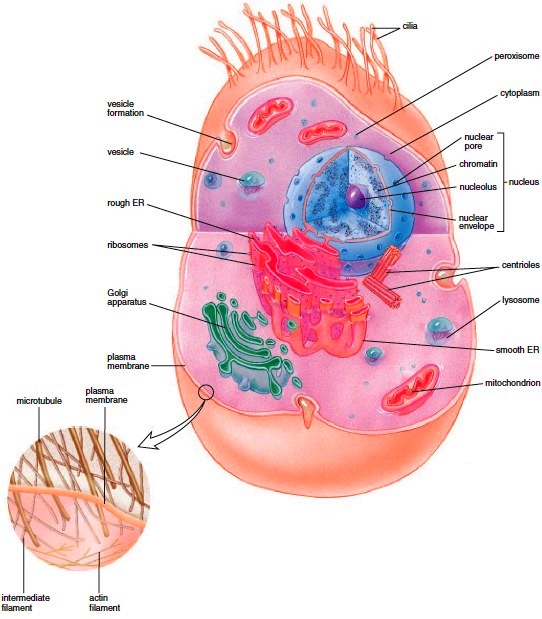
Figure 3.1 A generalized cell, with a blowup of the cytoskeleton.
Cells also have a cytoskeleton, a network of interconnected filaments and microtubules in the cytoplasm. The name cytoskeleton is convenient in that it allows us to compare the cytoskeleton to our bones and muscles. Bones and muscles give us structure and produce movement. Similarly, the elements of the cytoskeleton maintain cell shape and allow the cell and its contents to move. Some cells move by using cilia and flagella, which are made up of microtubules.

The Plasma Membrane
Our cells are surrounded by an outer plasma membrane. The plasma membrane separates the inside of the cell, termed the cytoplasm, from the outside. Plasma membrane integrity is necessary to the life of the cell. The plasma membrane is a phospholipid bilayer with attached or embedded proteins. The phospholipid molecule has a polar head and nonpolar tails (Fig. 3.2a). Because the polar heads are charged, they are hydrophilic (water-loving) and face outward, where they are likely to encounter a watery environment. The nonpolar tails are hydrophobic (waterfearing) and face inward, where there is no water. When phospholipids are placed in water, they naturally form a spherical bilayer because of the chemical properties of the heads and the tails. At body temperature, the phospholipid bilayer is a liquid; it has the consistency of olive oil, and the proteins are able to change their positions by moving laterally. The fluid-mosaic model, a working description of membrane structure, suggests that the protein molecules have a changing pattern (form a mosaic) within the fluid phospholipid bilayer (Fig. 3.2b). Our plasma membranes also contain a substantial number of cholesterol molecules. These molecules lend stability to the phospholipid bilayer and prevent a drastic decrease in fluidity at low temperatures. Short chains of sugars are attached to the outer surfaces of some protein and lipid molecules (called glycoproteins and glycolipids, respectively). These carbohydrate chains, specific to each cell, mark the cell as belonging to a particular individual and account for such characteristics as blood type or why a patient’s system sometimes rejects an organ transplant. Some glycoproteins have a special configuration that allows them to act as a receptor for a chemical messenger such as a hormone. Some plasma membrane proteins form channels through which certain substances can enter cells, while others are carriers involved in the passage of molecules through the membrane.
Our cells are surrounded by an outer plasma membrane. The plasma membrane separates the inside of the cell, termed the cytoplasm, from the outside. Plasma membrane integrity is necessary to the life of the cell. The plasma membrane is a phospholipid bilayer with attached or embedded proteins. The phospholipid molecule has a polar head and nonpolar tails (Fig. 3.2a). Because the polar heads are charged, they are hydrophilic (water-loving) and face outward, where they are likely to encounter a watery environment. The nonpolar tails are hydrophobic (waterfearing) and face inward, where there is no water. When phospholipids are placed in water, they naturally form a spherical bilayer because of the chemical properties of the heads and the tails. At body temperature, the phospholipid bilayer is a liquid; it has the consistency of olive oil, and the proteins are able to change their positions by moving laterally. The fluid-mosaic model, a working description of membrane structure, suggests that the protein molecules have a changing pattern (form a mosaic) within the fluid phospholipid bilayer (Fig. 3.2b). Our plasma membranes also contain a substantial number of cholesterol molecules. These molecules lend stability to the phospholipid bilayer and prevent a drastic decrease in fluidity at low temperatures. Short chains of sugars are attached to the outer surfaces of some protein and lipid molecules (called glycoproteins and glycolipids, respectively). These carbohydrate chains, specific to each cell, mark the cell as belonging to a particular individual and account for such characteristics as blood type or why a patient’s system sometimes rejects an organ transplant. Some glycoproteins have a special configuration that allows them to act as a receptor for a chemical messenger such as a hormone. Some plasma membrane proteins form channels through which certain substances can enter cells, while others are carriers involved in the passage of molecules through the membrane.
Figure 3.2 Fluid-mosaic model of the plasma membrane. a. In the phospholipid bilayer, the polar (hydrophilic) heads project outward and the nonpolar (hydrophobic) tails project inward. b. Proteins are embedded in the membrane. Glycoproteins have attached carbohydrate chains as do glycolipids.
The Nucleus
The nucleus is a prominent structure in human cells. The nucleus is of primary importance because it stores the genetic information that determines the characteristics of the body’s cells and their metabolic functioning. Every cell contains a copy of genetic information, but each cell type has certain genes turned on, and others turned off. Activated DNA, with messenger RNA (mRNA) acting as an intermediary, controls protein synthesis. The proteins of a cell determine its structure and the functions it can perform. When you look at the nucleus, even in an electron micrograph, you cannot see DNA molecules, but you can see chromatin (Fig. 3.3). Chemical analysis shows that chromatin contains DNA and much protein, as well as some RNA. Chromatin undergoes coiling into rodlike structures called chromosomes just before the cell divides. Chromatin is immersed in a semifluid medium called nucleoplasm. Most likely, too, when you look at an electron micrograph of a nucleus (Fig. 3.3), you will see one or more regions that look darker than the rest of the chromatin. These are nucleoli (sing., nucleolus) where another type of RNA, called ribosomal RNA (rRNA), is produced and where rRNA joins with proteins to form the subunits of ribosomes. (Ribosomes are small bodies in the cytoplasm that contain rRNA and proteins.) The nucleus is separated from the cytoplasm by a double membrane known as the nuclear envelope, which is continuous with the endoplasmic reticulum. The nuclear envelope has nuclear pores of sufficient size to permit the passage of proteins into the nucleus and ribosomal subunits out of the nucleus.
The nucleus is a prominent structure in human cells. The nucleus is of primary importance because it stores the genetic information that determines the characteristics of the body’s cells and their metabolic functioning. Every cell contains a copy of genetic information, but each cell type has certain genes turned on, and others turned off. Activated DNA, with messenger RNA (mRNA) acting as an intermediary, controls protein synthesis. The proteins of a cell determine its structure and the functions it can perform. When you look at the nucleus, even in an electron micrograph, you cannot see DNA molecules, but you can see chromatin (Fig. 3.3). Chemical analysis shows that chromatin contains DNA and much protein, as well as some RNA. Chromatin undergoes coiling into rodlike structures called chromosomes just before the cell divides. Chromatin is immersed in a semifluid medium called nucleoplasm. Most likely, too, when you look at an electron micrograph of a nucleus (Fig. 3.3), you will see one or more regions that look darker than the rest of the chromatin. These are nucleoli (sing., nucleolus) where another type of RNA, called ribosomal RNA (rRNA), is produced and where rRNA joins with proteins to form the subunits of ribosomes. (Ribosomes are small bodies in the cytoplasm that contain rRNA and proteins.) The nucleus is separated from the cytoplasm by a double membrane known as the nuclear envelope, which is continuous with the endoplasmic reticulum. The nuclear envelope has nuclear pores of sufficient size to permit the passage of proteins into the nucleus and ribosomal subunits out of the nucleus.

Ribosomes
Ribosomes are composed of two subunits, one large and one small. Each subunit has its own mix of proteins and rRNA. Protein synthesis occurs at the ribosomes. Ribosomes are found free within the cytoplasm either singly or in groups called polyribosomes. Ribosomes are often attached to the endoplasmic reticulum, a membranous system of saccules and channels discussed next (Fig. 3.4). Proteins synthesized by cytoplasmic ribosomes are used inside the cell for various purposes. Those produced by ribosomes attached to endoplasmic reticulum may eventually be secreted from the cell.
Ribosomes are composed of two subunits, one large and one small. Each subunit has its own mix of proteins and rRNA. Protein synthesis occurs at the ribosomes. Ribosomes are found free within the cytoplasm either singly or in groups called polyribosomes. Ribosomes are often attached to the endoplasmic reticulum, a membranous system of saccules and channels discussed next (Fig. 3.4). Proteins synthesized by cytoplasmic ribosomes are used inside the cell for various purposes. Those produced by ribosomes attached to endoplasmic reticulum may eventually be secreted from the cell.

Figure 3.3 The nucleus. The nuclear envelope with pores (arrows) surrounds the chromatin. Chromatin has a special region called the nucleolus, where rRNA is produced and ribosomal subunits are assembled.
Figure 3.4 Rough endoplasmic reticulum is studded with ribosomes where protein synthesis occurs. Smooth endoplasmic reticulum, which has no attached ribosomes, produces lipids and often has other functions as well in particular cells.
Contacts: lubopitno_bg@abv.bg www.encyclopedia.lubopitko-bg.com Corporation. All rights reserved.
DON'T FORGET - KNOWLEDGE IS EVERYTHING!