Central Nervous System
The CNS, consisting of the brain and spinal cord, is composed of gray matter and white matter. Gray matter is gray because it contains cell bodies and short, nonmyelinated fibers. White matter is white because it contains myelinated axons that run together in bundles called tracts.
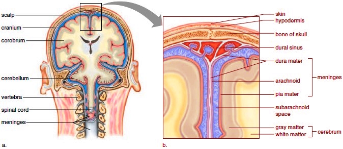
Figure 8.6 Meninges. a. Meninges are protective membranes that enclose the brain and spinal cord. b. The meninges include three layers: the dura mater, the arachnoid, and the pia mater.

Meninges and Cerebrospinal Fluid
Both the spinal cord and the brain are wrapped in protective membranes known as meninges (sing., meninx). The outer meninx, the dura mater, is tough, white, fibrous connective tissue that lies next to the skull and vertebrae. The dural sinuses collect venous blood before it returns to the cardiovascular system. Bleeding into the space between the dura mater and bone is called an epidural hematoma. The presence of blood between the dura mater and the next meninx, the arachnoid, is called a subdural hematoma. The arachnoid consists of weblike connective tissue with thin strands that attach it to the pia mater, the deepest meninx. The subarachnoid space is filled with cerebrospinal fluid, a clear tissue fluid that forms a protective cushion around and within the CNS. The pia mater is very thin and closely follows the contours of the brain and spinal cord (Fig. 8.6). Cerebrospinal fluid is stored within the central canal of the spinal cord and in the brain’s ventricles, which are interconnecting chambers that also produce cerebrospinal fluid. Normally, any excess cerebrospinal fluid drains away into the cardiovascular system. However, blockages can occur. In an infant, the brain can enlarge due to cerebrospinal fluid accumulation, resulting in a condition called hydrocephalus (“water on the brain”).
Both the spinal cord and the brain are wrapped in protective membranes known as meninges (sing., meninx). The outer meninx, the dura mater, is tough, white, fibrous connective tissue that lies next to the skull and vertebrae. The dural sinuses collect venous blood before it returns to the cardiovascular system. Bleeding into the space between the dura mater and bone is called an epidural hematoma. The presence of blood between the dura mater and the next meninx, the arachnoid, is called a subdural hematoma. The arachnoid consists of weblike connective tissue with thin strands that attach it to the pia mater, the deepest meninx. The subarachnoid space is filled with cerebrospinal fluid, a clear tissue fluid that forms a protective cushion around and within the CNS. The pia mater is very thin and closely follows the contours of the brain and spinal cord (Fig. 8.6). Cerebrospinal fluid is stored within the central canal of the spinal cord and in the brain’s ventricles, which are interconnecting chambers that also produce cerebrospinal fluid. Normally, any excess cerebrospinal fluid drains away into the cardiovascular system. However, blockages can occur. In an infant, the brain can enlarge due to cerebrospinal fluid accumulation, resulting in a condition called hydrocephalus (“water on the brain”).
The Spinal Cord
The spinal cord is a cylinder of nervous tissue that begins at the base of the brain and extends through a large opening in the skull called the foramen magnum. The spinal cord is protected by the vertebral column, which is composed of individual vertebrae. The cord passes through the vertebral canal formed by openings in the vertebrae. It ends at the first lumbar vertebra.
Structure of the Spinal Cord
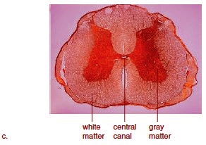
Figure 8.7a shows how an individual vertebra protects the spinal cord. The spinal nerves extend from the cord between the vertebrae. Intervertebral disks separate the vertebrae, and if a disk slips a bit and presses on the spinal cord, pain results. A cross section of the spinal cord shows a central canal, gray matter, and white matter (Fig. 8.7b,c). The central canal contains cerebrospinal fluid, as do the meninges that protect the spinal cord. The gray matter is centrally located and shaped like the letter H. Portions of sensory neurons and motor neurons are found there, as are interneurons that communicate with these two types of neurons. The posterior (dorsal) root of a spinal nerve contains sensory fibers entering the gray matter, and the anterior (ventral) root of a spinal nerve contains motor fibers exiting the gray matter. The posterior and anterior roots join, forming a spinal nerve that leaves the vertebral canal. Spinal nerves are a part of the PNS. The white matter of the spinal cord contains ascending tracts taking information to the brain (primarily located posteriorly) and descending tracts taking information from the brain (primarily located anteriorly). Because the tracts cross just after they enter and exit the brain, the left side of the brain controls the right side of the body, and the right side of the brain controls the left side of the body.
Figure 8.7a shows how an individual vertebra protects the spinal cord. The spinal nerves extend from the cord between the vertebrae. Intervertebral disks separate the vertebrae, and if a disk slips a bit and presses on the spinal cord, pain results. A cross section of the spinal cord shows a central canal, gray matter, and white matter (Fig. 8.7b,c). The central canal contains cerebrospinal fluid, as do the meninges that protect the spinal cord. The gray matter is centrally located and shaped like the letter H. Portions of sensory neurons and motor neurons are found there, as are interneurons that communicate with these two types of neurons. The posterior (dorsal) root of a spinal nerve contains sensory fibers entering the gray matter, and the anterior (ventral) root of a spinal nerve contains motor fibers exiting the gray matter. The posterior and anterior roots join, forming a spinal nerve that leaves the vertebral canal. Spinal nerves are a part of the PNS. The white matter of the spinal cord contains ascending tracts taking information to the brain (primarily located posteriorly) and descending tracts taking information from the brain (primarily located anteriorly). Because the tracts cross just after they enter and exit the brain, the left side of the brain controls the right side of the body, and the right side of the brain controls the left side of the body.

Figure 8.7 Spinal cord. a. The spinal cord passes through the vertebral canal formed by the vertebrae.It gives off spinal nerves that project through openings between the vertebrae.

Figure 8.7 Spinal cord. b. The spinal cord has a central canal filled with cerebrospinal fluid, graymatter in an H-shaped configuration, and white matter elsewhere.

Functions of the Spinal Cord
The spinal cord provides a means of communication between the brain and the peripheral nerves that leave the cord. When someone touches your hand, sensory receptors generate nerve impulses that pass through sensory fibers to the spinal cord and up one of several ascending tracts to a sensory area of the brain. When you voluntarily move your limbs, motor impulses originating in the brain pass down one of several descending tracts to the spinal cord and out to your muscles by way of motor fibers. The spinal cord is also the center for thousands of reflex arcs: A stimulus causes sensory receptors to generate nerve impulses that travel in sensory neurons to the spinal cord. Interneurons integrate the incoming data and relay signals to motor neurons. A response to the stimulus occurs when motor axons cause skeletal muscles to contract. Each interneuron in the spinal cord has synapses with many other neurons, and therefore they send signals to several other interneurons in addition to motor neurons.
The spinal cord provides a means of communication between the brain and the peripheral nerves that leave the cord. When someone touches your hand, sensory receptors generate nerve impulses that pass through sensory fibers to the spinal cord and up one of several ascending tracts to a sensory area of the brain. When you voluntarily move your limbs, motor impulses originating in the brain pass down one of several descending tracts to the spinal cord and out to your muscles by way of motor fibers. The spinal cord is also the center for thousands of reflex arcs: A stimulus causes sensory receptors to generate nerve impulses that travel in sensory neurons to the spinal cord. Interneurons integrate the incoming data and relay signals to motor neurons. A response to the stimulus occurs when motor axons cause skeletal muscles to contract. Each interneuron in the spinal cord has synapses with many other neurons, and therefore they send signals to several other interneurons in addition to motor neurons.
Figure 8.7 Spinal cord. c. Photomicrograph of a cross section of the spinal cord.
Contacts: lubopitno_bg@abv.bg www.encyclopedia.lubopitko-bg.com Corporation. All rights reserved.
DON'T FORGET - KNOWLEDGE IS EVERYTHING!