Circulatory Routes
Blood vessels belong to either the pulmonary circuit or the systemic circuit. The path of blood through the pulmonary circuit can be traced as follows: Blood from all regions of the body first collects in the right atrium and then passes into the right ventricle, which pumps it into the pulmonary trunk. The pulmonary trunk divides into the pulmonary arteries, which in turn divide into the arterioles of the lungs. The arterioles then take blood to the pulmonary capillaries, where carbon dioxide and oxygen are exchanged. The blood then enters the pulmonary venules and flows through the pulmonary veins back to the left atrium. Because the blood in the pulmonary arteries is O2-poor but the blood in the pulmonary veins is O2-rich, it is not correct to say that all arteries carry blood that is high in oxygen and that all veins carry blood that is low in oxygen. In fact, just the reverse is true in the pulmonary circuit.
The systemic circuit includes all of the other arteries and veins of the body. The largest artery in the systemic circuit is the aorta, and the largest veins are the superior vena cava and inferior vena cava. The superior vena cava collects blood from the head, chest, and arms, and the inferior vena cava collects blood from the lower body regions. Both venae cavae enter the right atrium. The aorta and venae cavae are the major pathways for blood in the systemic system.
The path of systemic blood to any organ in the body begins in the left ventricle, which pumps blood into the aorta. Branches from the aorta go to the major body regions and organs. Tracing the path of blood to any organ in the body requires mentioning only the aorta, the proper branch of the aorta, the organ, and the returning vein to the vena cava. In many instances, the artery and vein that serve the same organ have the same name. For example, the path of blood to and from the kidneys is: left ventricle; aorta; renal artery; arterioles, capillaries, venules; renal vein; inferior vena cava; right atrium. In the systemic circuit, unlike the pulmonary circuit, arteries contain O2-rich blood and appear bright red, while veins contain O2-poor blood and appear dark maroon.
The Major Systemic Arteries
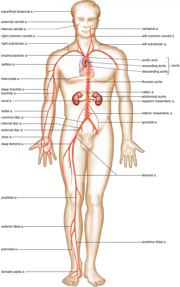
After the aorta leaves the heart, it divides into the ascending aorta, the aortic arch, and the descending aorta (Fig. 12.18). The left and right coronary arteries, which supply blood to the heart, branch off the ascending aorta (Table. 12.1).
Three major arteries branch off the aortic arch: the brachiocephalic artery, the left common carotid artery, and the left subclavian artery. The brachiocephalic artery divides into the right common carotid and the right subclavian arteries. These blood vessels serve the head (right and left common carotids) and arms (right and left subclavians).
The descending aorta is divided into the thoracic aorta, which branches off to the organs within the thoracic cavity, and the abdominal aorta, which branches off to the organs in the abdominal cavity. The descending aorta ends when it divides into the common iliac arteries that branch into the internal iliac artery and the external iliac artery. The internal iliac artery serves the pelvic organs, and the external iliac artery serves the legs. These and other arteries are shown in Figure 12.18.


Figure 12.18 Major arteries (a.) of the body.

The Major Systemic Veins
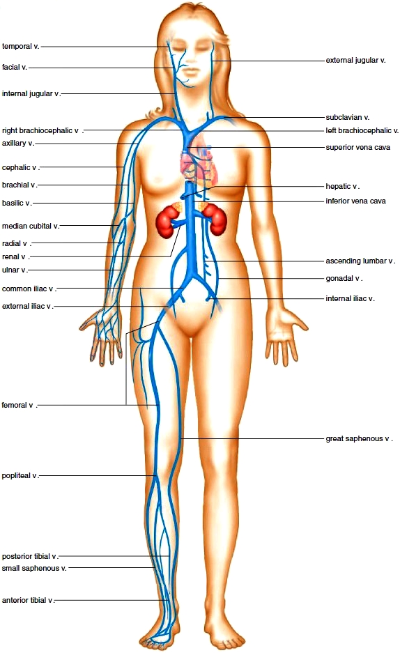
Figure 12.19 shows the major veins of the body. The external and internal jugular veins drain blood from the brain, head, and neck. An external jugular vein enters a subclavian vein that, along with an internal jugular vein, enters a brachiocephalic vein. Right and left brachiocephalic veins merge, giving rise to the superior vena cava.
In the abdominal cavity, the hepatic portal vein receives blood from the abdominal viscera and enters the liver. Emerging from the liver, the hepatic veins enter the inferior vena cava.
In the pelvic region, veins from the various organs enter the internal iliac veins, while the veins from the legs enter the external iliac veins. The internal and external iliac veins become the common iliac veins that merge, forming the inferior vena cava. Table 12.2 lists the principal veins that enter the venae cavae.
Special Systemic Circulations
Hepatic Portal System
The hepatic portal system (Fig. 12.20) carries blood from the stomach, intestines, and other organs to the liver. The term portal system is used to describe the following unique pattern of circulation:
capillaries - vein - capillaries - vein
Capillaries of the digestive tract drain into the superior mesenteric vein and the splenic vein, which join to form the hepatic portal vein. The gastric veins empty directly into the hepatic portal vein. The hepatic portal vein carries blood to capillaries in the liver. The hepatic capillaries allow nutrients and wastes to diffuse into liver cells for further processing. Then, hepatic capillaries join to form venules that enter a hepatic vein. The hepatic veins enter the inferior vena cava.
In addition to receiving venous blood from the intestine, the liver also receives arterial blood via the hepatic artery. The hepatic artery is not a part of the hepatic portal system.
Hypothalamus-Hypophyseal Portal System
The body has other portal systems. For example, the vascular link between the hypothalamus and the anterior pituitary through which the hypothalamus sends hypothalamicreleasing hormones to the anterior pituitary is a portal system.

Figure 12.20 Hepatic portal system. This system provides venous drainage of the digestive organs and takes venous blood to the liver. (v. = vein).

Blood Supply to the Brain
The brain is supplied with O2-rich blood by the anterior and posterior cerebral arteries and the carotid arteries. These arteries give off branches. that join to form the cerebral arterial circle (circle of Willis), a vascular route in the region of the pituitary gland (Fig. 12.21). Because the blood vessels form a circle, alternate routes are available for bringing arterial blood to the brain and thus supplying the brain with oxygen. The presence of the cerebral arterial circle also equalizes blood pressure in the brain’s blood supply.

Figure 12.21 Cerebral arterial circle. The arteries that supply blood to the brain form the cerebral arterial circle (circle ofWillis). (a. = artery).
Figure 12.19 Major veins (v.) of the body.
Fetal Circulation
As Figure 12.22 shows, the fetus has four circulatory features that are not present in adult circulation:
1. The foramen ovale, or oval window, is an opening between the two atria. This window is covered by a flap of tissue that acts as a valve.
2. The ductus arteriosus, or arterial duct, is a connection between the pulmonary artery and the aorta.
3. The umbilical arteries and vein are vessels that travel to and from the placenta, leaving waste and receiving nutrients.
4. The ductus venosus, or venous duct, is a connection between the umbilical vein and the inferior vena cava.
All of these features can be related to the fact that the fetus does not use its lungs for gas exchange, since it receives oxygen and nutrients from the mother’s blood at the placenta. During development, the lungs receive only enough blood to supply their developmental need for oxygen and nutrients. The path of blood in the fetus can be traced, beginning from the right atrium (Fig. 12.22). Most of the blood that enters the right atrium passes directly into the left atrium by way of the foramen ovale because the blood pressure in the right atrium is somewhat greater than that in the left atrium. The rest of the fetal blood entering the right atrium passes into the right ventricle and out through the pulmonary trunk. However, because of the ductus arteriosus, most pulmonary trunk blood passes directly into the aortic arch. Notice that, whatever route blood takes, most of it reaches the aortic arch instead of the pulmonary circuit vessels. Blood within the aorta travels to the various branches, including the iliac arteries, which connect to the umbilical arteries leading to the placenta. Exchange between maternal and fetal blood takes place at the placenta. Blood in the umbilical arteries is O2-poor, but blood in the umbilical vein, which travels from the placenta, is O2-rich. The umbilical vein enters the ductus venosus, which passes directly through the liver. The ductus venosus then joins with the inferior vena cava, a vessel that contains O2-poor blood. The vena cava returns this mixture to the right atrium.
Changes at Birth Sectioning and tying the umbilical cord permanently separates the newborn from the placenta. The first breath inflates the lungs and oxygen enters the blood at the lungs instead of the placenta. O2-rich blood returning from the lungs to the left side of the heart usually causes a flap on the left side of the interatrial septum to close the foramen ovale. What remains is a depression called the fossa ovalis. Incomplete closure occurs in nearly one out of four individuals, but even so, blood rarely passes from the right atrium to the left atrium because either the opening is small or it closes when the atria contract. In a small number of cases, the passage of O2-poor blood from the right side to the left side of the heart is sufficient to cause cyanosis, a bluish cast to the skin. This condition can now be corrected by openheart surgery.
The fetal blood vessels and shunts constrict and become fibrous connective tissue called ligamentums in all cases except the distal portions of the umbilical arteries, which become the medial umbilical ligaments. Regardless, these structures run between internal organs. For example, the ligamentum teres attaches the umbilicus to the liver.
Figure 12.22 Fetal circulation. Arrows indicate the direction of blood flow. The lungs are not functional in the fetus. The blood passes directly from the right atrium to the left atrium via the foramen ovale or from the right ventricle to the aorta via the pulmonary trunk and ductus venosus. The umbilical arteries take fetal blood to the placenta where exchange of molecules between fetal and maternal blood takes place. Oxygen and nutrient molecules diffuse into the fetal blood, and carbon dioxide and urea diffuse from the fetal blood. The umbilical vein returns blood from the placenta to the fetus.

Contacts: lubopitno_bg@abv.bg www.encyclopedia.lubopitko-bg.com Corporation. All rights reserved.
DON'T FORGET - KNOWLEDGE IS EVERYTHING!