Extracellular Junctions, Glands, and Membranes
Extracellular Junctions
The cells of a tissue can function in a coordinated manner when the plasma membranes of adjoining cells interact. The junctions that occur between cells help cells function as a tissue. A tight junction forms an impermeable barrier because adjacent plasma membrane proteins actually join, producing a zipperlike fastening (Fig. 4.15a). In the small intestine, gastric juices stay out of the body, and in the kidneys, the urine stays within kidney tubules because epithelial cells are joined by tight junctions. A gap junction forms when two adjacent plasma membrane channels join (Fig. 4.15b). This lends strength, but it also allows ions, sugars, and small molecules to pass between the two cells. Gap junctions in heart and smooth muscle ensure synchronized contraction. In an adhesion junction (desmosome), the adjacent plasma membranes do not touch but are held together by extracellular filaments firmly attached to cytoplasmic plaques, composed of dense protein material (Fig. 4.15c).
Glands
A gland consists of one or more cells that produce and secrete a product. Most glands are composed primarily of epithelium in which the cells secrete their product by exocytosis.

Figure 4.15 Extracellular junctions. Tissues are held together by (a) tight junctions that are impermeable; (b) gap junctions that allow materials to pass from cell to cell; and (c) adhesion junctions that allow tissues to stretch.
During secretion, the contents of a vesicle are released when the vesicle fuses with the plasma membrane. The mucus-secreting goblet cells within the columnar epithelium lining the digestive tract are single cells (see Fig. 4.3). Glands with ducts that secrete their product onto the outer surface (e.g., sweat glands and mammary glands) or into a cavity (e.g., pancreas) are called exocrine glands. Ducts can be simple or compound, as illustrated in Figure 4.16. Glands that no longer have a duct are appropriately known as the ductless glands, or endocrine glands. Endocrine glands (e.g., pituitary gland and thyroid) secrete their products internally so they are transported by the bloodstream. Endocrine glands produce hormones that help promote homeostasis. Each type of hormone influences the metabolism of a particular target organ or cells. Glands are composed of epithelial tissue, but they are supported by connective tissue, as are other epithelial tissues.
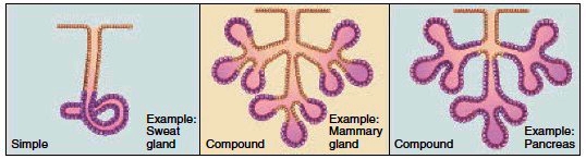
Figure 4.16 Multicellular exocrine glands. Exocrine glands have ducts that can be simple or compound. Compound glands vary according to the placement of secretory portions.
Membranes
Membranes line the internal spaces of organs and tubes that open to the outside, and they also line the body cavities.
Mucous Membranes
Mucous membranes line the interior walls of the organs and tubes that open to the outside of the body, such as those of the digestive, respiratory, urinary, and reproductive systems. These membranes consist of an epithelium overlying a layer of loose connective tissue. The epithelium contains goblet cells that secrete mucus. The mucus secreted by mucous membranes ordinarily protects interior walls from invasion by bacteria and viruses; for example, more mucus is secreted when a person has a cold, resulting in a “runny nose.” In addition, mucus usually protects the walls of the stomach and small intestine from digestive juices, but this protection breaks down when a person develops an ulcer.
Serous Membranes
Serous membranes line cavities, including the thoracic and abdominopelvic cavities, and cover internal organs such as the intestines. The term parietal refers to the wall of the body cavity, while the term visceral pertains to the internal organs. Therefore, parietal membranes line the interior of the thoracic and abdominopelvic cavities, and visceral membranes cover the organs. Serous membranes consist of a layer of simple squamous epithelium overlying a layer of loose connective tissue. They secrete a watery fluid that keeps the membranes lubricated. Serous membranes support the internal organs and tend to compartmentalize the large thoracic and abdominopelvic cavities. This helps hinder the spread of any infection. In the thorax, the pleura are serous membranes that form a double layer around the lungs. The parietal pleura lines the inside of the thoracic wall, while the visceral pleura adheres to the surface of the lungs. Similarly a double-layered serous membrane is a part of the pericardium, a covering for the heart. The peritoneum is the serous membranes within the abdomen. The parietal peritoneum lines the abdominopelvic wall, and the visceral peritoneum covers the organs. In between the organs, the visceral peritoneum comes together to form a double-layered mesentery that supports these organs.
Synovial Membranes
Synovial membranes line freely movable joint cavities and are composed of connective tissues. They secrete synovial fluid into the joint cavity; this fluid lubricates the ends of the bones so that they can move freely. In rheumatoid arthritis, the synovial membrane becomes inflamed and grows thicker. Fibrous tissue then invades the joint and may eventually become bony so that the bones of the joint are no longer capable of moving.
Meninges
The meninges are membranes found within the posterior cavity (see Fig. 1.5). They are composed only of connective tissue and serve as a protective covering for the brain and spinal cord. Meningitis is a life-threatening infection of the meninges.
Cutaneous Membrane
The cutaneous membrane, or skin, forms the outer covering of the body. It consists of an outer portion of keratinized stratified squamous epithelium attached to a thick underlying layer of dense irregular connective tissue.
Contacts: lubopitno_bg@abv.bg www.encyclopedia.lubopitko-bg.com Corporation. All rights reserved.
DON'T FORGET - KNOWLEDGE IS EVERYTHING!