Imaging the Body
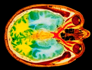
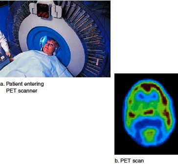
Imaging the body for diagnosis of disease is based on chemical properties of subatomic particles. For example, X rays, which are produced when high-speed electrons strike a heavy metal, have long been used to image body parts. Dense structures such as bone absorb X rays well and show up as light areas; soft tissues absorb X rays to a lesser extent and show up as dark areas on photographic film. During CAT (computerized axial tomography) scans, X rays are sent through the body at various angles, and a computer uses the X-ray information to form a series of cross sections (Fig. 1B). CAT scanning has reduced the need for exploratory surgery and can guide the surgeon in visualizing complex body structures during surgical procedures. PET (positron emission tomography) is a variation on CT scanning. Radioactively labeled substances are injected into the body; metabolically active tissues tend to take up these substances and then emit gamma rays. A computer uses the gamma-ray information to again generate cross-sectional images of the body, but this time, the image indicates metabolic activity, not structure (see Fig. 2.3). PET scanning is used to diagnose brain disorders, such as a brain tumor, Alzheimer disease, epilepsy, or stroke. During MRI (magnetic resonance imaging), the patient lies in a massive, hollow, cylindrical magnet and is exposed to short bursts of a powerful magnetic field. This causes the protons in the nuclei of hydrogen atoms to align. Then, when exposed to strong radio waves, the protons move out of alignment and produce signals.

Figure 1B CAT (computerized axial tomography).

Tissues with many hydrogen atoms (such as fat) show up as bright areas, while tissues with few hydrogen atoms (such as bone) appear black. This is the opposite of an X ray, which is why MRI is more useful than an X ray for imaging soft tissues. However, many people cannot undergo MRI, because the magnetic field can actually pull a metal object out of the body, such as a tooth filling, a prosthesis, or a pacemaker!
Figure 2.3 Use of radiation to study the brain. After the administration of radioactively labeled glucose, a PET scan reveals which portions of the brain are most active.

Contacts: lubopitno_bg@abv.bg www.encyclopedia.lubopitko-bg.com Corporation. All rights reserved.
DON'T FORGET - KNOWLEDGE IS EVERYTHING!