Lymphoid Tissue
Lymphoid tissue is distributed throughout the body and makes up the specialized organs of the lymphatic system. The lymph nodes have already been described relative to describing lymphatic circulation, but these tissues and other components of the lymphatic system are discussed in greater detail in the next page.
Lymph Nodes
The lymph nodes, as noted, are designed to filter the lymph once it is drained from the tissues (Fig. 12-5). They are also sites where lymphocytes of the immune system multiply and work to combat foreign organisms. The lymph nodes are small, rounded masses varying from pinhead size to as long as 2.5 cm (1 inch). Each has a fibrous connective tissue capsule from which partitions (trabeculae) extend into the substance of the node. At various points in the node’s surface, afferent lymphatic vessels pierce the capsule to carry lymph into the node. An indented area called the hilum is the exit point for efferent lymphatic vessels carrying lymph out of the node. At this region, other structures, including blood vessels and nerves, connect with the node. Each node is subdivided into lymph-filled spaces (sinuses) and cords of lymphatic tissue.
Pulplike nodules in the outer region, or cortex, have germinal centers where certain immune lymphocytes multiply. The inner region, the medulla, has populations of immune cells, including lymphocytes and macrophages (phagocytes) along open channels that lead into the efferent vessels. Lymph nodes are seldom isolated. As a rule, they are massed together in groups, varying in number from 2 or 3 to well over 100. Some of these groups are placed deeply, whereas others are superficial. The main groups include the following:

Figure 12-5 Structure of a lymph node. (A) Arrows indicate the flow of lymph through the node. (B) Section of a lymph node as seen under the microscope
* Cervical nodes, located in the neck in deep and superficial groups, drain various parts of the head and neck. They often become enlarged during upper respiratory infections.
* Axillary nodes, located in the axillae (armpits), may become enlarged after infections of the upper extremities and the breasts. Cancer cells from the breasts often metastasize (spread) to the axillary nodes.
* Tracheobronchial nodes are found near the trachea and around the larger bronchial tubes. In people living in highly polluted areas, these nodes become so filled with carbon particles that they are solid black masses resembling pieces of coal.
* Mesenteric nodes are found between the two layers of peritoneum that form the mesentery There are some 100 to 150 of these nodes.
* Inguinal nodes, located in the groin region, receive lymph drainage from the lower extremities and from the external genital organs. When they become enlarged, they are often referred to as buboes, from which bubonic plague got its name.
The Spleen
The spleen is an organ that contains lymphoid tissue designed to filter blood. It is located in the superior left hypochondriac region of the abdomen, high up under the dome of the diaphragm, and normally is protected by the lower part of the rib cage (Fig. 12-6). The spleen is a soft, purplish, and somewhat flattened organ, measuring approximately 12.5 to 16 cm (5 to 6 inches) long and 5 to 7.5 cm (2 to 3 inches) wide. The capsule of the spleen, as well as its framework, is more elastic than that of the lymph nodes. It contains involuntary muscle, which enables the splenic capsule to contract and also to withstand some swelling.
Considering its size, the spleen has an unusually large blood supply. The organ is filled with a soft pulp that filters the blood. It also harbors phagocytes and lymphocytes, which are active in immunity.
The spleen is classified as part of the lymphatic system because it contains prominent masses of lymphoid tissue. However, it has wider functions than other lymphatic structures, including the following:
* Cleansing the blood of impurities and cellular debris by filtration and phagocytosis.
* Destroying old, worn-out red blood cells. The iron and other breakdown products of hemoglobin are carried to the liver by the hepatic portal system to be reused or eliminated from the body.
* Producing red blood cells before birth.
* Serving as a reservoir for blood, which can be returned to the bloodstream in case of hemorrhage or other emergency.
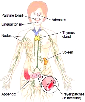
Figure 12-6 Location of lymphoid tissue.
Splenectomy, or surgical removal of the spleen, is usually a well tolerated procedure. Although the spleen is the largest unit of lymphoid tissue in the body, other lymphoid tissues can take over its functions. The human body has thousands of lymphoid units, and the loss of any one unit or group ordinarily is not a threat to life.
The Thymus
Because of its appearance under a microscope, the thymus, located in the superior thorax beneath the sternum, traditionally has been considered part of the lymphoid system (see Fig. 12-6). Recent studies, however, suggest that this structure has a much wider function than other lymphoid tissue. It appears that the thymus plays a key role in immune system development before birth and during the first few months of infancy. Certain lymphocytes must mature in the thymus gland before they can perform their functions in the immune system. These T cells (T lymphocytes) develop under the effects of the thymus gland hormone called thymosin, which also promotes lymphocyte growth and activity in lymphoid tissue throughout the body. Removal of the thymus causes a decrease in the production of T cells, as well as a decrease in the size of the spleen and of lymph nodes throughout the body. The thymus is most active during early life. After puberty, the tissue undergoes changes; it shrinks in size and is replaced by connective tissue and fat.
The Tonsils
The tonsils are masses of lymphoid tissue located in the vicinity of the pharynx (throat) where they remove contaminants from materials that are inhaled or swallowed (Fig. 12-7). The tonsils have deep grooves lined with lymphatic nodules. Lymphocytes attack pathogens trapped in these grooves. The tonsils are located in three areas:
* The palatine tonsils are oval bodies located at each side of the soft palate. These are generally what is meant when one refers to the tonsils.
* The single pharyngeal tonsil is commonly referred to as the adenoids (from a general term that means “gland-like”). It is located behind the nose on the posterior wall of the upper pharynx.
* The lingual tonsils are little mounds of lymphoid tissue at the back of the tongue. Any of these tonsils may become so loaded with bacteria that they become reservoirs for repeated infections and their removal is advisable. In children, a slight enlargement of any of them is not an indication for surgery, however, because all lymphoid tissue masses tend to be larger in childhood. A physician must determine whether these masses are abnormally enlarged, taking the patient’s age into account, because the tonsils function in immunity during early childhood. The surgery to remove the palatine tonsils is a tonsillectomy; an adenoidectomy is removal of the adenoids. Often these two procedures are done together and abbreviated as T & A.
Other Lymphoid Tissue
The appendix is a fingerlike tube of lymphatic tissue, measuring about approximately 8 cm (3 in.) long, and is attached, or “appended” to the first portion of the large intestine (see Fig. 12-6). Like the tonsils, it seems to be noticed only when it becomes infected, causing appendicitis. The appendix may, however, figure in the development of immunity, as do the tonsils. In the mucous membranes lining portions of the digestive, respiratory, and urogenital tracts there are areas of lymphatic tissue that help destroy outside contaminants.

Figure 12-7 Location of the tonsils. All are in the vicinity of the pharynx (throat).
By means of phagocytosis and production of antibodies, substances that counteract infectious agents, this mucosal-associated lymphoid tissue, or MALT, prevents microorganisms from invading deeper tissues. Peyer patches are part of the MALT system. These clusters of lymphatic nodules are located in the mucous membranes lining the small intestine’s distal portion. Peyer patches, along with the tonsils and appendix, are included in the specific network known as GALT, or gut-associated lymphoid tissue. All of these lymphatic tissues associated with mucous membranes are now recognized as an important first barrier against invading microorganisms.
Contacts: lubopitno_bg@abv.bg www.encyclopedia.lubopitko-bg.com Corporation. All rights reserved.
DON'T FORGET - KNOWLEDGE IS EVERYTHING!