Muscles of the Upper Extremities
Muscles of the upper extremities include the muscles that determine the position of the shoulder, the anterior and posterior muscles that move the arm, and the muscles that move the forearm and hand.
Muscles That Move the Shoulder and Arm
The position of the shoulder depends to a large extent on the degree of contraction of the trapezius, a triangular muscle that covers the posterior neck and extends across the posterior shoulder to insert on the clavicle and scapula (Fig. 4-10). The trapezius muscles enable one to raise the shoulders and pull them back. The upper portion of each trapezius can also extend the head and turn it from side to side. The latissimus dorsi is the wide muscle of the back and lateral trunk. It originates from the vertebral spine in the middle and lower back and covers the lower half of the thoracic region, forming the posterior portion of the axilla (armpit). The fibers of each muscle converge to a tendon that inserts on the humerus. The latissimus dorsi powerfully extends the arm, bringing it down forcibly as, for example, in swimming. A large pectoralis major is located on either side of the superior part of the chest (see Fig. 4-9). This muscle arises from the sternum, the upper ribs, and the clavicle and forms the anterior “wall” of the armpit, or axilla; it inserts on the superior part of the humerus. The pectoralis major flexes and adducts the arm, pulling it across the chest.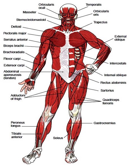
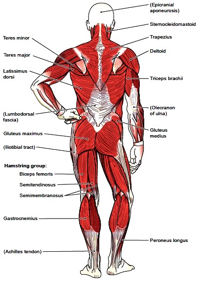
Figure 4-9 Superficial muscles, anterior view. Associated structure is labeled in parentheses.
Figure 4-10 Superficial muscles, posterior view. Associated structures are labeled in parentheses.
The serratus anterior is below the axilla, on the lateral part of the chest. It originates on the upper eight or nine ribs on the lateral and anterior thorax and inserts in the scapula on the side toward the vertebrae. The serratus anterior moves the scapula forward when, for example, one is pushing something. It also aids in raising the arm above the horizontal level. The deltoid covers the shoulder joint and is responsible for the roundness of the upper part of the arm just inferior to the shoulder (see Figs. 4-9 and 4-10). This muscle is named for its triangular shape, which resembles the Greek letter delta. The deltoid is often used as an injection site. Arising from the shoulder girdle (clavicle and scapula), the deltoid fibers converge to insert on the lateral surface of the humerus. Contraction of this muscle abducts the arm, raising it laterally to the horizontal position. The shoulder joint allows for a very wide range of movement. This freedom of movement is possible because the humerus fits into a shallow socket, the glenoid cavity of the scapula. This joint requires the support of four deep muscles and their tendons, which compose the rotator cuff. The four muscles are the supraspinatus, infraspinatus, teres minor, and subscapularis, known together as SITS, based on the first letters of their names. In certain activities, such as swinging a golf club, playing tennis, or pitching a baseball, the muscles of the rotator cuff may be injured, even torn, and may require surgery for repair.
Muscles That Move the Forearm and Hand
The biceps brachii, located at the anterior arm along the humerus, is the muscle you usually display when you want to “flex your muscles” to show your strength (Fig. 4-12 A). It inserts on the radius and flexes the forearm. It is a supinator of the hand. Another flexor of the forearm at the elbow is the brachioradialis, a prominent muscle of the forearm that originates at the distal end of the humerus and inserts on the distal radius. The triceps brachii, located on the posterior of the arm, inserts on the olecranon of the ulna (Fig. 4-12 B). It is used to straighten the arm, as in lowering a weight from an arm curl. It is also important in pushing because it converts the arm and forearm into a sturdy rod. Most of the muscles that move the hand and fingers originate from the radius and the ulna (see Fig. 4-12). Some of them insert on the carpal bones of the wrist, whereas others have long tendons that cross the wrist and insert on bones of the hand and the fingers. The flexor carpi and the extensor carpi muscles are responsible for many movements of the hand. Muscles that produce finger movements are the several flexor digitorum and the extensor digitorum muscles. The names of these muscles may include bones they are near, their action, or their length, for example, longus for long and brevis for short. Special groups of muscles in the fleshy parts of the hand are responsible for the intricate movements that can be performed with the thumb and the fingers. The thumb’s freedom of movement has been one of the most useful capacities of humans.Muscles of the Trunk
The muscles of the trunk include the muscles involved in breathing, the thin muscle layers of the abdomen, and the muscles of the pelvic floor. The following discussion also includes the deep muscles of the back that support and move the vertebral column.
Muscles of Respiration
The most important muscle involved in the act of breathing is the diaphragm. This dome-shaped muscle forms the partition between the thoracic cavity superiorly and the abdominal cavity inferiorly (Fig. 4-13). When the diaphragm contracts, the central dome-shaped portion is pulled downward, thus enlarging the thoracic cavity from top to bottom. The intercostal muscles are attached to and fill the spaces between the ribs. The external and internal intercostals run at angles in opposite directions. Contraction of the intercostal muscles serves to elevate the ribs, thus enlarging the thoracic cavity from side to side and from anterior to posterior.
Figure 4-12 Muscles that move the forearm and hand.

Figure 4-13 Muscles of respiration. Associated structures are also shown.
Muscles of the Abdomen and Pelvis
The wall of the abdomen has three layers of muscle that extend from the back (dorsally) and around the sides (laterally) to the front (ventrally) (Fig. 4-14). They are the external oblique on the outside, the internal oblique in the middle, and the transversus abdominis, the innermost. The connective tissue from these muscles extends forward and encloses the vertical rectus abdominis of the anterior abdominal wall. The fibers of these muscles, as well as their connective tissue extensions (aponeuroses), run in different directions, resembling the layers in plywood and resulting in a strong abdominal wall. The midline meeting of the aponeuroses forms a whitish area called the linea alba, which is an important landmark on the abdomen. It extends from the tip of the sternum to the pubic joint. These four pairs of abdominal muscles act together to protect the internal organs and compress the abdominal cavity, as in coughing, emptying the bladder (urination) and bowel (defecation), sneezing, vomiting, and childbirth (labor). The two oblique muscles and the rectus abdominis help bend the trunk forward and sideways. The pelvic floor, or perineum, has its own form of diaphragm, shaped somewhat like a shallow dish. One of the principal muscles of this pelvic diaphragm is the levator ani, which acts on the rectum and thus aids in defecation. The superficial and deep muscles of the female perineum are shown in Figure 4-15 along with some associated structures.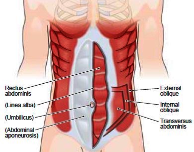
Figure 4-14 Muscles of the abdominal wall. Surface tissue is removed on the right side to show deeper muscles. Associated structures are labeled in parentheses.

Figure 4-15 Muscles of the female perineum (pelvic floor). Associated structures are labeled in parentheses.
Deep Muscles of the Back
The deep muscles of the back, which act on the vertebral column itself, are thick vertical masses that lie under the trapezius and latissimus dorsi. The erector spinae muscles make up a large group located between the sacrum and the skull. These muscles extend the spine and maintain the vertebral column in an erect posture. The muscles can be strained in lifting heavy objects if the spine is flexed while lifting. One should bend at the hip and knee instead and use the thigh and buttock muscles to help in lifting. Deeper muscles in the lumbar area extend the vertebral column in that region. These deep muscles of the back are not shown in the illustrations.
Contacts: lubopitno_bg@abv.bg www.encyclopedia.lubopitko-bg.com Corporation. All rights reserved.
DON'T FORGET - KNOWLEDGE IS EVERYTHING!