Peripheral Nervous System
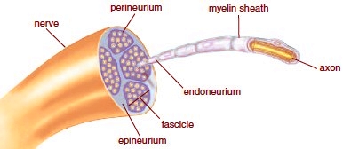
The peripheral nervous system (PNS) lies outside the central nervous system and is composed of nerves and ganglia. Nerves are bundles of myelinated axons. Ganglia (sing., ganglion) are swellings associated with nerves that contain collections of cell bodies. As with muscles, connective tissue separates axons at various levels of organization:

The PNS is subdivided into the somatic system and the autonomic system. The somatic system serves the skin, skeletal muscles, and tendons. It includes nerves that take sensory information from external sensory receptors to the CNS and motor commands away from the CNS to the skeletal muscles. The autonomic system, with a few exceptions, regulates the activity of cardiac and smooth muscles and glands.
Types of Nerves
The cranial nerves are attached to the brain, and the spinal nerves are attached to the spinal cord.
Cranial Nerves
Humans have 12 pairs of cranial nerves (Table 8.1). By convention, the pairs of cranial nerves are referred to by roman numerals (Fig. 8.11a). Most of the cranial nerves belong to the somatic system. Some of these are sensory nerves-that is, they contain only sensory fibers; some are motor nerves, containing only motor fibers; and others are mixed nerves, so called because they contain both sensory and motor fibers. Cranial nerves are largely concerned with the head, neck, and facial regions of the body. However, the vagus nerve (X), which has branches to most of the internal organs, is a part of the autonomic system.

Figure 8.11 Cranial and spinal nerves. a. Ventral surface of the brain showing the attachment of the 12 pairs of cranial nerves. b. Cross section of the spinal cord, showing 3 pairs of spinal nerves. Each spinal nerve has a posterior root and an anterior root that join shortly beyond the cord.

Spinal Nerves
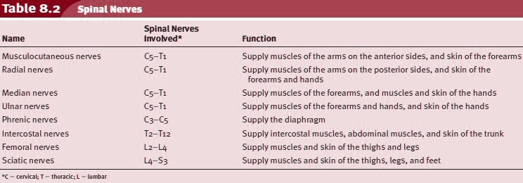
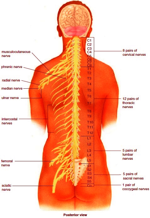
Humans have 31 pairs of spinal nerves; one of each pair is on either side of the spinal cord (Fig. 8.11b). The spinal nerves are grouped as shown in Table 8.2 because they are at either the cervical, thoracic, or lumbar regions of the vertebral column. The spinal nerves are designated according to their location in relation to the vertebrae because each passes through an intervertebral foramen as it leaves the spinal cord. This organizational principle is illustrated in Figure 8.12.
Humans have 31 pairs of spinal nerves; one of each pair is on either side of the spinal cord (Fig. 8.11b). The spinal nerves are grouped as shown in Table 8.2 because they are at either the cervical, thoracic, or lumbar regions of the vertebral column. The spinal nerves are designated according to their location in relation to the vertebrae because each passes through an intervertebral foramen as it leaves the spinal cord. This organizational principle is illustrated in Figure 8.12.


Figure 8.12 Spinal nerves. The number and kinds of spinal nerves are given on the right. The location ofmajor peripheral nerves is given on the left. Table 8.2 lists the functions of these nerves.
Many spinal nerves carry fibers that belong to either the somatic or the autonomic system. However, the spinal nerves are called mixed nerves because they contain both sensory fibers that conduct impulses to the spinal cord from sensory receptors and motor fibers that conduct impulses away from the cord to effectors. The sensory fibers enter the cord via the posterior root, and the motor fibers exit by way of the anterior root. The cell body of a sensory neuron is in a posterior (dorsal)-root ganglion. Each spinal nerve serves the particular region of the body in which it is located.
Many actions in the somatic nervous system are voluntary, and these always originate in the cerebral cortex, as when we decide to move a limb. Other actions in the somatic nervous system are due to reflexes, automatic involuntary responses to changes occurring inside or outside the body. A reflex occurs quickly, without our even having to think about it. Some reflexes, called cranial reflexes, involve the brain, as when we automatically blink our eyes when an object nears the eye suddenly. Figure 8.13 illustrates the path of a reflex within the somatic nervous system that involves only the spinal cord (called a spinal reflex). If your hand touches a sharp pin, a sensory receptor in the skin generates nerve impulses that move along a sensory fiber through the posteriorroot ganglia toward the spinal cord. Sensory neurons enter the cord posteriorly and pass signals on to many interneurons. Some of these interneurons synapse with motor neurons whose short dendrites and cell bodies are in the spinal cord. Nerve impulses travel along a motor fiber to an effector, which brings about a response to the stimulus. In this case, the effector is a skeletal muscle, which contracts so that you withdraw your hand from the pin.
Various other reactions are also possible-you will most likely look at the pin, wince, and cry out in pain. This whole series of responses occurs because certain interneurons carry nerve impulses to the brain via tracts in the spinal cord and brain. The brain makes you aware of the stimulus and directs your other reactions to it. You don’t feel pain until the brain receives the information and interprets it. Reflexes are essential to homeostasis. They keep the internal organs functioning within normal bounds and protect the body from external harm.
Reflexes can also be used to determine if the nervous system is reacting properly. Two of these types of reflexes are:
knee-jerk reflex (patellar reflex), initiated by striking the patellar ligament just below the patella. The response is contraction of the quadriceps femoris muscles, which causes the lower leg to extend;
ankle-jerk reflex, initiated by tapping the Achilles tendon just above its insertion on the calcaneus. The response is plantar flexion due to contraction of the gastrocnemius and soleus muscles.
Some reflexes are important for avoiding injury, but the knee-jerk and ankle-jerk reflexes are important for normal physiological functions. For example, the knee-jerk reflex helps a person stand erect. If the knee begins to bend slightly when a person stands still, the quadriceps femoris is stretched, and the leg straightens.
Somatic Nervous System
Many actions in the somatic nervous system are voluntary, and these always originate in the cerebral cortex, as when we decide to move a limb. Other actions in the somatic nervous system are due to reflexes, automatic involuntary responses to changes occurring inside or outside the body. A reflex occurs quickly, without our even having to think about it. Some reflexes, called cranial reflexes, involve the brain, as when we automatically blink our eyes when an object nears the eye suddenly. Figure 8.13 illustrates the path of a reflex within the somatic nervous system that involves only the spinal cord (called a spinal reflex). If your hand touches a sharp pin, a sensory receptor in the skin generates nerve impulses that move along a sensory fiber through the posteriorroot ganglia toward the spinal cord. Sensory neurons enter the cord posteriorly and pass signals on to many interneurons. Some of these interneurons synapse with motor neurons whose short dendrites and cell bodies are in the spinal cord. Nerve impulses travel along a motor fiber to an effector, which brings about a response to the stimulus. In this case, the effector is a skeletal muscle, which contracts so that you withdraw your hand from the pin.
Various other reactions are also possible-you will most likely look at the pin, wince, and cry out in pain. This whole series of responses occurs because certain interneurons carry nerve impulses to the brain via tracts in the spinal cord and brain. The brain makes you aware of the stimulus and directs your other reactions to it. You don’t feel pain until the brain receives the information and interprets it. Reflexes are essential to homeostasis. They keep the internal organs functioning within normal bounds and protect the body from external harm.
Reflexes can also be used to determine if the nervous system is reacting properly. Two of these types of reflexes are:
knee-jerk reflex (patellar reflex), initiated by striking the patellar ligament just below the patella. The response is contraction of the quadriceps femoris muscles, which causes the lower leg to extend;
ankle-jerk reflex, initiated by tapping the Achilles tendon just above its insertion on the calcaneus. The response is plantar flexion due to contraction of the gastrocnemius and soleus muscles.
Some reflexes are important for avoiding injury, but the knee-jerk and ankle-jerk reflexes are important for normal physiological functions. For example, the knee-jerk reflex helps a person stand erect. If the knee begins to bend slightly when a person stands still, the quadriceps femoris is stretched, and the leg straightens.
Figure 8.13 A reflex arc showing the path of a spinal reflex. A stimulus (e.g., a pinprick) causes sensory receptors in the skin to generate nerve impulses that travel in sensory axons to the spinal cord. Interneurons integrate data from sensory neurons and then relay signals to motor neurons. Motor axons convey nerve impulses from the spinal cord to a skeletal muscle, which contracts. Movement of the hand away from the pin is the response to the stimulus.

Contacts: lubopitno_bg@abv.bg www.encyclopedia.lubopitko-bg.com Corporation. All rights reserved.
DON'T FORGET - KNOWLEDGE IS EVERYTHING!