Protein Synthesis
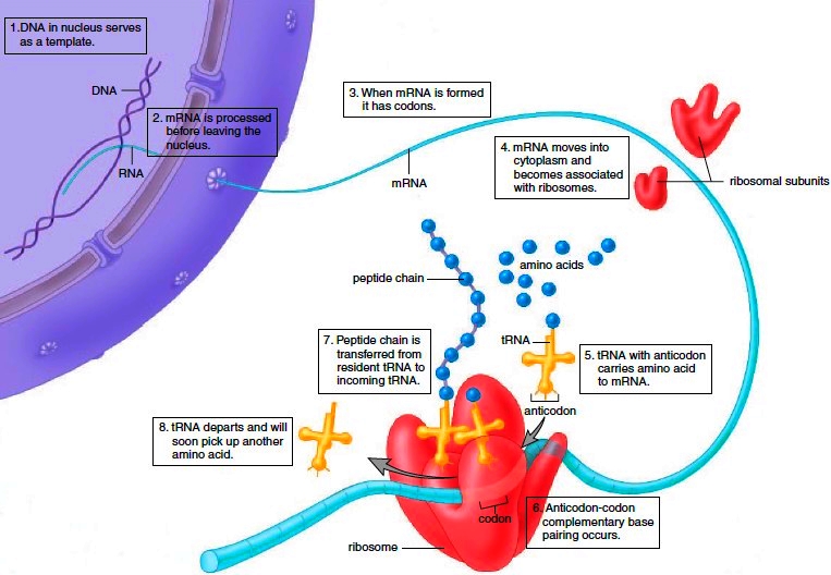
DNA not only serves as a template for its own replication, but is also a template for RNA formation. Protein synthesis requires two steps, called transcription and translation. During transcription, an mRNA molecule is produced, and during translation, this mRNA specifies the order of amino acids in a particular polypeptide (Fig. 3.13). A gene (i.e., DNA) contains coded information for the sequence of amino acids in a particular polypeptide. The code is a triplet code: Every three bases in DNA (and therefore in mRNA) stands for a particular amino acid.
Transcription and Translation
During transcription, complementary RNA nucleotides from an RNA nucleotide pool in the nucleus pair with the DNA nucleotides of one strand. The RNA nucleotides are joined by an enzyme called RNA polymerase, and an mRNA molecule results. Therefore, when mRNA forms, it has a sequence of bases complementary to DNA. A sequence of three bases that are complementary to the DNA triplet code is a codon. Translation requires several enzymes and two other types of RNA: transfer RNA and ribosomal RNA. Transfer RNA (tRNA) molecules bring amino acids to the ribosomes, which are composed of ribosomal RNA (rRNA) and protein. There is at least one tRNA molecule for each of the 20 amino acids found in proteins. The amino acid binds to one end of the molecule, and the entire complex is designated as tRNA-amino acid. At the other end of each tRNA molecule is a specific anticodon, a group of three bases that is complementary to an mRNA codon. A tRNA molecule comes to the ribosome, where its anticodon pairs with an mRNA codon. For example, if the codon is ACC, then the anticodon is UGG and the amino acid is threonine.
Figure 3.13 Protein synthesis. The two steps required for protein synthesis are transcription, which occurs in the nucleus, and translation, which occurs in the cytoplasm at the ribosomes.
(The codes for each of the 20 amino acids are known.) Notice that the order of the codons of the mRNA determines the order that tRNA-amino acids come to a ribosome, and therefore the final sequence of amino acids in a polypeptide.

Events During the Mitotic Stage
The mitotic stage of the cell cycle consists of mitosis and cytokinesis. By the end of interphase (Fig. 3.14, upper left), the centrioles have doubled and the chromosomes are becoming visible. Each chromosome is duplicated-it is composed of two chromatids held together at a centromere. As an aid in describing the events of mitosis, the process is divided into four phases: prophase, metaphase, anaphase, and telophase (Fig. 3.14). The parental cell is the cell that divides, and the daughter cells are the cells that result.
Prophase
Several events occur during prophase that visibly indicate the cell is about to divide. The two pairs of centrioles outside the nucleus begin moving away from each other toward opposite ends of the nucleus. Spindle fibers appear between the separating centriole pairs, the nuclear envelope begins to fragment, and the nucleolus begins to disappear. The chromosomes are now fully visible. Although humans have 46 chromosomes, only four are shown in Figure 3.14 for ease in following the phases of mitosis. Spindle fibers attach to the centromeres as the chromosomes continue to shorten and thicken. During prophase, chromosomes are randomly placed in the nucleus.
Structure of the Spindle At the end of prophase, a cell has a fully formed spindle. A spindle has poles, asters, and fibers. The asters are arrays of short microtubules that radiate from the poles, and the fibers are bundles of microtubules that stretch between the poles. Centrioles are located in centrosomes, which are believed to organize the spindle.
Figure 3.14 The late interphase cell and the mitotic stage of the cell cycle. Although humans have 46 chromosomes, only four are shown here for convenience. The blue chromosomes were originally inherited from a father, and the red were originally inherited from a mother.

Metaphase
During metaphase, the nuclear envelope is fragmented, and the spindle occupies the region formerly occupied by the nucleus. The chromosomes are now at the equator (center) of the spindle. Metaphase is characterized by a fully formed spindle, and the chromosomes, each with two sister chromatids, are aligned at the equator (Fig. 3.15).
Anaphase
At the start of anaphase, the sister chromatids separate. Once separated, the chromatids are called chromosomes. Separation of the sister chromatids ensures that each cell receives a copy of each type of chromosome and thereby has a full complement of genes. During anaphase, the daughter chromosomes move to the poles of the spindle. Anaphase is characterized by the movement of chromosomes toward each pole.
Function of the Spindle The spindle brings about chromosome movement. Two types of spindle fibers are involved in the movement of chromosomes during anaphase. One type extends from the poles to the equator of the spindle; there, they overlap. As mitosis proceeds, these fibers increase in length, and this helps push the chromosomes apart. The chromosomes themselves are attached to other spindle fibers that simply extend from their centromeres to the poles. These fibers get shorter and shorter as the chromosomes move toward the poles. Therefore, they pull the chromosomes apart. Spindle fibers, as stated earlier, are composed of microtubules. Microtubules can assemble and disassemble by the addition or subtraction of tubulin (protein) subunits.

Figure 3.15 Micrographs of mitosis occurring in a whitefish embryo.
This is what enables spindle fibers to lengthen and shorten, and it ultimately causes the movement of the chromosomes.
Telophase and Cytokinesis
Telophase begins when the chromosomes arrive at the poles. During telophase, the chromosomes become indistinct chromatin again. The spindle disappears as nucleoli appear, and nuclear envelope components reassemble in each cell. Telophase is characterized by the presence of two daughter nuclei.
Cytokinesis is division of the cytoplasm and organelles. In human cells, a slight indentation called a cleavage furrow passes around the circumference of the cell. Actin filaments form a contractile ring, and as the ring gets smaller and smaller, the cleavage furrow pinches the cell in half. As a result, each cell becomes enclosed by its own plasma membrane.
Importance of Mitosis
Because of mitosis, each cell in our body is genetically identical, meaning that it has the same number and kinds of chromosomes. Mitosis is important to the growth and repair of multicellular organisms. When a baby develops in the mother’s womb, mitosis occurs as a component of growth. As a wound heals, mitosis occurs, and the damage is repaired.
Contacts: lubopitno_bg@abv.bg www.encyclopedia.lubopitko-bg.com Corporation. All rights reserved.
DON'T FORGET - KNOWLEDGE IS EVERYTHING!