The Cerebral Hemispheres
Each cerebral hemisphere is divided into four visible lobes named for the overlying cranial bones. These are the frontal, parietal, temporal, and occipital lobes (Fig. 6-6). In addition, there is a small fifth lobe deep within each hemisphere that cannot be seen from the surface. Not much is known about this lobe, which is called the insula. The outer nervous tissue of the cerebral hemispheres is gray matter that makes up the cerebral cortex (see Fig. 6-3). This thin layer of gray matter (2-4 mm thick) is the most highly evolved portion of the brain and is responsible for conscious thought, reasoning, and abstract mental functions. Specific functions are localized in the cortex of the different lobes, as described in greater detail later. The cortex is arranged in folds forming elevated portions known as gyri, singular gyrus. These raised areas are separated by shallow grooves called sulci, singular sulcus (Fig. 6-7). Although there are many sulci, the following two are especially important landmarks:
* The central sulcus, which lies between the frontal and parietal lobes of each hemisphere at right angles to the longitudinal fissure (see Figs. 6-2 and 6-6);
* The lateral sulcus, which curves along the side of each hemisphere and separates the temporal lobe from the frontal and parietal lobes (see Fig. 6-6).
Internally, the cerebral hemispheres are made largely of white matter and a few islands of gray matter. The white matter consists of myelinated fibers that connect the cortical areas with each other and with other parts of the nervous system. Basal nuclei, also called basal ganglia, are masses of gray matter located deep within each cerebral hemisphere. These groups of neurons work with the cerebral cortex to regulate body movement and the muscles of facial expression. The neurons of the basal nuclei secrete the neurotransmitter dopamine. The corpus callosum is an important band of white matter located at the bottom of the longitudinal fissure (see Fig. 6-1). This band is a bridge between the right and left hemispheres, permitting impulses to cross from one side of the brain to the other. The internal capsule is a compact band of myelinated fibers that carries impulses between the cerebral hemispheres and the brain stem. The vertical fibers that make up the internal capsule travel between the thalamus and some of the basal nuclei on each side and then radiate toward the cerebral cortex.
Figure 6-6 External surface of the brain, lateral view. The lobes and surface features of the cerebrum are visible.


Figure 6-7 Section of the cerebrum. Labels point out surface features, the cerebral cortex, and the white matter.
Functions of the Cerebral Cortex
It is within the cerebral cortex, the layer of gray matter that forms the surface of each cerebral hemisphere, that impulses are received and analyzed. These activities form the basis of knowledge. The brain “stores” information,
much of which can be recalled on demand by means of the phenomenon called memory. It is in the cerebral cortex that thought processes such as association, judgment, and discrimination take place. Conscious deliberation and voluntary actions also arise from the cerebral cortex. Although the various brain areas act in coordination to produce behavior, particular functions are localized in the cortex of each lobe (Fig. 6-8). Some of these are described below:
* The frontal lobe, which is relatively larger in humans than in any other organism, lies anterior to the central sulcus. The gyrus just anterior to the central sulcus in this lobe contains a primary motor area, which provides conscious control of skeletal muscles. Note that the more detailed the action, the greater the amount of cortical tissue involved (Fig. 6-9). The frontal lobe also contains two areas important in speech (the speech centers are discussed later).

Figure 6-8 Functional areas of the cerebral cortex.
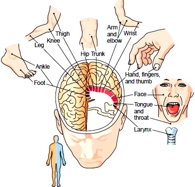
Figure 6-9 Motor areas of the cerebral cortex (frontal lobe). The amount of cortex involved in control of a body part is proportional to the degree of coordination needed in movement. The small figure indicates that control is contralateral. The right hemisphere controls the left side of the body and the left hemisphere controls the right side of the body.
* The parietal lobe occupies the superior part of each hemisphere and lies posterior to the central sulcus. The gyrus just behind the central sulcus in this lobe contains the primary sensory area, where impulses from the skin, such as touch, pain, and temperature, are interpreted. The estimation of distances, sizes, and shapes also takes place here. As with the motor cortex, the greater the intensity of sensation from a particular area, the tongue or fingers, for example, the more area of the cortex is involved.
* The temporal lobe lies inferior to the lateral sulcus and folds under the hemisphere on each side. This lobe contains the auditory area for receiving and interpreting impulses from the ear. The olfactory area, concerned with the sense of smell, is located in the medial part of the temporal lobe; it is stimulated by impulses arising from receptors in the nose.
* The occipital lobe lies posterior to the parietal lobe and extends over the cerebellum. The visual area of this lobe contains the visual receiving area and the visual association area for interpreting impulses arising from the retina of the eye.
The ability to communicate by written and verbal means is an interesting example of the way in which areas of the cerebral cortex are interrelated (see Fig. 6-8). The development and use of these areas are closely connected with the process of learning.
* The auditory areas lie in the temporal lobe. One of these areas, the auditory receiving area, detects sound impulses transmitted from the environment, whereas the surrounding area, the auditory association area, interprets the sounds. Another region of the auditory cortex, the speech comprehension area, or Wernicke area, functions in speech recognition and the meaning of words. Someone who suffers damage in this region of the brain, as by a stroke, will have difficulty in understanding the meaning of speech. The beginnings of language are learned by hearing; thus, the auditory areas for understanding sounds are near the auditory receiving area of the cortex. Babies often appear to understand what is being said long before they do any talking themselves. It is usually several years before children learn to read or write words.
* The motor areas for spoken and written communication lie anterior to the most inferior part of the frontal lobe’s motor cortex. The speech muscles in the tongue, the soft palate, and the larynx are controlled here, in a region named the motor speech area, or Broca area (see Fig. 6-8). A person who suffers damage to this area may have difficulty in producing speech (motor aphasia). Similarly, the written speech center lies anterior to the cortical area that controls the arm and hand muscles. The ability to write words is usually one of the last phases in the development of learning words and their meanings.
* The visual areas of the occipital lobe’s cortex are also involved in communication. Here, visual images of language are received. The visual area that lies anterior to the receiving cortex then interprets these visual impulses as words. The ability to read with understanding also develops in this area. You might see writing in the Japanese language, for example, but this would involve only the visual receiving area in the occipital lobe unless you could also understand the words. There is a functional relation among areas of the brain. Many neurons must work together to enable a person to receive, interpret, and respond to verbal and written messages as well as to touch (tactile stimulus) and other sensory stimuli.
Memory is the mental faculty for recalling ideas. In the initial stage of the memory process, sensory signals (e.g., visual, auditory) are retained for a very short time, perhaps only fractions of a second. Nevertheless, they can be used for further processing. Short-term memory refers to the retention of bits of information for a few seconds or perhaps a few minutes, after which the information is lost unless reinforced. Long-term memory refers to the storage of information that can be recalled at a later time. There is a tendency for a memory to become more fixed the more often a person repeats the remembered experience; thus, short-term memory signals can lead to long-term memories. Furthermore, the more often a memory is recalled, the more indelible it becomes; such a memory can be so deeply fixed in the brain that it can be recalled immediately. Careful anatomic studies have shown that tiny extensions called fibrils form at the synapses in the cerebral cortex, enabling impulses to travel more easily from one neuron to another. The number of these fibrils increases with age. Physiologic studies show that rehearsal (repetition) of the same information again and again accelerates and potentiates the degree of transfer of short-term memory into long-term memory. A person who is wide awake memorizes far better than does a person who is in a state of mental fatigue. It has also been noted that the brain is able to organize information so that new ideas are stored in the same areas in which similar ones had been stored before.
* The temporal lobe lies inferior to the lateral sulcus and folds under the hemisphere on each side. This lobe contains the auditory area for receiving and interpreting impulses from the ear. The olfactory area, concerned with the sense of smell, is located in the medial part of the temporal lobe; it is stimulated by impulses arising from receptors in the nose.
* The occipital lobe lies posterior to the parietal lobe and extends over the cerebellum. The visual area of this lobe contains the visual receiving area and the visual association area for interpreting impulses arising from the retina of the eye.
Communication Areas
The ability to communicate by written and verbal means is an interesting example of the way in which areas of the cerebral cortex are interrelated (see Fig. 6-8). The development and use of these areas are closely connected with the process of learning.
* The auditory areas lie in the temporal lobe. One of these areas, the auditory receiving area, detects sound impulses transmitted from the environment, whereas the surrounding area, the auditory association area, interprets the sounds. Another region of the auditory cortex, the speech comprehension area, or Wernicke area, functions in speech recognition and the meaning of words. Someone who suffers damage in this region of the brain, as by a stroke, will have difficulty in understanding the meaning of speech. The beginnings of language are learned by hearing; thus, the auditory areas for understanding sounds are near the auditory receiving area of the cortex. Babies often appear to understand what is being said long before they do any talking themselves. It is usually several years before children learn to read or write words.
* The motor areas for spoken and written communication lie anterior to the most inferior part of the frontal lobe’s motor cortex. The speech muscles in the tongue, the soft palate, and the larynx are controlled here, in a region named the motor speech area, or Broca area (see Fig. 6-8). A person who suffers damage to this area may have difficulty in producing speech (motor aphasia). Similarly, the written speech center lies anterior to the cortical area that controls the arm and hand muscles. The ability to write words is usually one of the last phases in the development of learning words and their meanings.
* The visual areas of the occipital lobe’s cortex are also involved in communication. Here, visual images of language are received. The visual area that lies anterior to the receiving cortex then interprets these visual impulses as words. The ability to read with understanding also develops in this area. You might see writing in the Japanese language, for example, but this would involve only the visual receiving area in the occipital lobe unless you could also understand the words. There is a functional relation among areas of the brain. Many neurons must work together to enable a person to receive, interpret, and respond to verbal and written messages as well as to touch (tactile stimulus) and other sensory stimuli.
Memory and the Learning Process
Memory is the mental faculty for recalling ideas. In the initial stage of the memory process, sensory signals (e.g., visual, auditory) are retained for a very short time, perhaps only fractions of a second. Nevertheless, they can be used for further processing. Short-term memory refers to the retention of bits of information for a few seconds or perhaps a few minutes, after which the information is lost unless reinforced. Long-term memory refers to the storage of information that can be recalled at a later time. There is a tendency for a memory to become more fixed the more often a person repeats the remembered experience; thus, short-term memory signals can lead to long-term memories. Furthermore, the more often a memory is recalled, the more indelible it becomes; such a memory can be so deeply fixed in the brain that it can be recalled immediately. Careful anatomic studies have shown that tiny extensions called fibrils form at the synapses in the cerebral cortex, enabling impulses to travel more easily from one neuron to another. The number of these fibrils increases with age. Physiologic studies show that rehearsal (repetition) of the same information again and again accelerates and potentiates the degree of transfer of short-term memory into long-term memory. A person who is wide awake memorizes far better than does a person who is in a state of mental fatigue. It has also been noted that the brain is able to organize information so that new ideas are stored in the same areas in which similar ones had been stored before.
Contacts: lubopitno_bg@abv.bg www.encyclopedia.lubopitko-bg.com Corporation. All rights reserved.
DON'T FORGET - KNOWLEDGE IS EVERYTHING!