Function of the Retina
The retina has a complex structure with multiple layers of cells (Fig. 7-4). The deepest layer is a pigmented layer just anterior to the choroid. Next are the rods and cones, the receptor cells of the eye, named for their shape. Anterior to the rods and cones are connecting neurons that carry impulses toward the optic nerve. The rods are highly sensitive to light and thus function in dim light, but they do not provide a sharp image. They are more numerous than the cones and are distributed more toward the periphery (anterior portion) of the retina. (If you visualize the retina as the inside of a bowl, the rods would be located toward the lip of the bowl). When you enter into dim light, such as a darkened movie theater, you cannot see for a short period. It is during this time that the rods are beginning to function, a change that is described as dark adaptation. When you are able to see again, images are blurred and appear only in shades of gray, because the rods are unable to differentiate colors. The cones function in bright light, are sensitive to color, and give sharp images. The cones are localized at the center of the retina, especially in a tiny depressed area near the optic nerve that is called the fovea centralis (Fig. 7-5; see also Fig. 7-3). (Note that fovea is a general term for a pit or depression.) Because this area contains the highest concentration of cones, it is the point of sharpest vision. The fovea is contained within a yellowish spot, the macula lutea, an area that may show degenerative changes with age. There are three types of cones, each sensitive to either red, green, or blue light. Color blindness results from a lack of retinal cones. People who completely lack cones are totally colorblind; those who lack one type of cone are partially color blind. This disorder, because of its pattern of inheritance, occurs almost exclusively in males. The rods and cones function by means of pigments that are sensitive to light. The rod pigment is rhodopsin, or visual purple. Vitamin A is needed for manufacture of these pigments. If a person is lacking in vitamin A, he or she may have difficulty seeing in dim light because there is too little light to activate the rods, a condition termed night blindness. Nerve impulses from the rods and cones flow into sensory neurons that eventually merge to form the optic nerve (cranial nerve II) at the eye’s posterior (see Figs. 7-3 and 7-5). The impulses travel to the visual center in the occipital cortex of the brain. When an ophthalmologist, a physician who specializes in treatment of the eye, examines the retina with an ophthalmoscope, he or she can see abnormalities in the retina and in the retinal blood vessels. Some of these changes may signal more widespread diseases that affect the eye, such as diabetes and high blood pressure (hypertension).

Figure 7-4 Structure of the retina. Rods and cones form a deep layer of the retina, near the choroid. Connecting neurons carry visual impulses toward the optic nerve.

Figure 7-5 The fundus (back) of the eye as seen through an ophthalmoscope.
Muscles of the Eye
Two groups of muscles are associated with the eye. Both groups are important in adjusting the eye so that a clear image can form on the retina.
The Extrinsic Muscles The voluntary muscles attached to the eyeball’s outer surface are the extrinsic muscles. The six ribbonlike extrinsic muscles connected with each eye originate on the bones of the orbit and insert on the surface of the sclera (Fig. 7-6). They are named for their location and the direction of the muscle fibers. These muscles pull on the eyeball in a coordinated fashion so that both eyes center on one visual field.
This process of convergence is necessary to the formation of a clear image on the retina.
Having the image come from a slightly different angle from each retina is believed to be important for three-dimensional (stereoscopic) vision, a characteristic of primates.

Figure 7-6 Extrinsic muscles of the eye. The medial rectus is not shown.

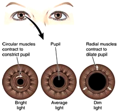
Figure 7-7 Function of the iris. In bright light, circular muscles contract and constrict the pupil, limiting the light that enters the eye. In dim light, the radial muscles contract and dilate the pupil, allowing more light to enter the eye.
The Intrinsic Muscles The involuntary muscles located within the eyeball are the intrinsic muscles. They form two circular structures within the eye, the iris and the ciliary muscle. The iris, the colored or pigmented part of the eye, is composed of two sets of muscle fibers that govern the size of the iris’s central opening, the pupil (Fig. 7-7). One set of fibers is arranged in a circular fashion, and the other set extends radially like the spokes of a wheel. The iris regulates the amount of light entering the eye. In bright light, the iris’s circular muscle fibers contract, reducing the size of the pupil. This narrowing is termed constriction. In contrast, in dim light, the radial muscles contract, pulling the opening outward and enlarging it. This enlargement of the pupil is known as dilation. The ciliary muscle is shaped somewhat like a flattened ring with a central hole the size of the outer edge of the iris. This muscle holds the lens in place by means of filaments, called suspensory ligaments, that project from the ciliary muscle to the edge of the lens around its entire circumference (Fig. 7-8). The ciliary muscle controls the shape of the lens to allow for vision at near and far distances. This process of accommodation occurs as follows. The light rays from a close object diverge (separate) more than do the light rays from a distant object (Fig. 7-9). Thus, when viewing something close, the lens must become more rounded to bend the light rays more and focus them on the retina. The ciliary muscle controls the shape of the lens. When this muscle is relaxed, tension on the suspensory ligaments keeps the lens in a more flattened shape. For close vision, the ciliary muscle contracts. This movement draws the ciliary ring forward and relaxes tension on the suspensory ligaments. The elastic lens then recoils and becomes thicker, in much the same way that a rubber band thickens when the pull on it is released.
When the ciliary muscle relaxes again, the lens flattens. These actions change the refractive power of the lens to accommodate for near and far vision. In young people, the lens is elastic, and therefore its thickness can be readily adjusted according to the need for near or distance vision. With aging, the lens loses elasticity and therefore its ability to accommodate for near vision. It becomes difficult to focus clearly on close objects. This condition is called presbyopia, which literally means “old eye.”

Figure 7-8 The ciliary muscle and lens (posterior view). Contraction of the ciliary muscle relaxes tension on the suspensory ligaments, allowing the lens to become more round for near vision.

Figure 7-9 Accommodation for near vision. When viewing a close object, the lens must become more rounded to focus light rays on the retina.
Nerve Supply to the Eye
Two sensory nerves supply the eye (Fig. 7-10):
* The optic nerve (cranial nerve II) carries visual impulses from the retinal rods and cones to the brain.
* The ophthalmic branch of the trigeminal nerve (cranial nerve V) carries impulses of pain, touch, and temperature from the eye and surrounding parts to the brain. The optic nerve arises from the retina a little toward the medial or nasal side of the eye. There are no retinal rods and cones in the area of the optic nerve. Consequently, no image can form on the retina at this point, which is known as the blind spot or optic disk (see Figs. 7-3 and 7-5). The optic nerve transmits impulses from the retina to the thalamus of the brain, from which they are directed to the occipital cortex. Note that the light rays passing through the eye are actually overrefracted (bent) so that an image falls on the retina upside down and backward (see Figs. 7-9). It is the job of the visual centers of the brain to reverse the images. Three nerves carry motor impulses to the eyeball muscles:
* The oculomotor nerve (cranial nerve III) is the largest; it supplies voluntary and involuntary motor impulses to all but two eye muscles.
* The trochlear nerve (cranial nerve IV) supplies the superior oblique extrinsic eye muscle (see Fig. 7-6).
* The abducens nerve (cranial nerve VI) supplies the lateral rectus extrinsic eye muscle.

Figure 7-10 Nerves of the eye.
The steps in vision are:
1. Light refracts.
2. The muscles of the iris adjust the pupil.
3. The ciliary muscle adjusts the lens (accommodation).
4. The extrinsic eye muscles produce convergence.
5. Light stimulates retinal receptor cells (rods and cones).
6. The optic nerve transmits impulses to the brain.
7. The occipital lobe cortex interprets the impulses.
Contacts: lubopitno_bg@abv.bg www.encyclopedia.lubopitko-bg.com Corporation. All rights reserved.
DON'T FORGET - KNOWLEDGE IS EVERYTHING!