Vertebral Column (Spine)
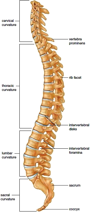
The vertebral column extends from the skull to the pelvis. It consists of a series of separate bones, the vertebrae, separated by pads of fibrocartilage called the intervertebral disks (Fig. 6.8). The vertebral column is located in the middorsal region and forms the vertical axis. The skull rests on the superior end of the vertebral column, which also supports the rib cage and serves as a point of attachment for the pelvic girdle. The vertebral column also protects the spinal cord, which passes through a vertebral canal formed by the vertebrae. The vertebrae are named according to their location: seven cervical (neck) vertebrae, twelve thoracic (chest) vertebrae, five lumbar (lower back) vertebrae, five sacral vertebrae fused to form the sacrum, and three to five coccygeal vertebrae fused into one coccyx.
When viewed from the side, the vertebral column has four normal curvatures, named for their location (Fig. 6.8). The cervical and lumbar curvatures are convex anteriorly, and the thoracic and sacral curvatures are concave anteriorly. In the fetus, the vertebral column has but one curve, and it is concave anteriorly. The cervical curve develops three to four months after birth, when the child begins to hold the head up. The lumbar curvature develops when a child begins to stand and walk, around one year of age. The curvatures of the vertebral column provide more support than a straight column would, and they also provide the balance needed to walk upright.
The curvatures of the vertebral column are subject to abnormalities. An abnormally exaggerated lumbar curvature is called lordosis, or “swayback.” People who are balancing a heavy midsection, such as pregnant women or men with “potbellies,” may have swayback.
An increased roundness of the thoracic curvature is kyphosis, or “hunchback.” This abnormality sometimes develops in older people. An abnormal lateral (side-to-side) curvature is called scoliosis. Occurring most often in the thoracic region, scoliosis is usually first seen during late childhood.

Figure 6.8 Curvatures of the spine. The vertebrae are named for their location in the body. Note the presence of the coccyx, also called the tailbone.
Intervertebral Disks
The fibrocartilaginous intervertebral disks located between the vertebrae act as a cushion. They prevent the vertebrae from grinding against one another and absorb shock caused by such movements as running, jumping, and even walking. The disks also allow motion between the vertebrae so that a person can bend forward, backward, and from side to side. Unfortunately, these disks become weakened with age, and can slip or even rupture (called a herniated disk). A damaged disk pressing against the spinal cord or the spinal nerves causes pain. Such a disk may need to be removed surgically. If a disk is removed, the vertebrae are fused together, limiting the body’s flexibility.
Vertebrae
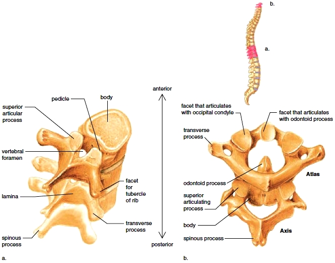
Figure 6.9a shows that a typical vertebra has an anteriorly placed body and a posteriorly placed vertebral arch. The vertebral arch forms the wall of a vertebral foramen (pl., foramina). The foramina become a canal through which the spinal cord passes. The vertebral spinous process (spine) occurs where two thin plates of bone called laminae meet. A transverse process is located where a pedicle joins a lamina. These processes serve for the attachment of muscles and ligaments. Articular processes (superior and inferior) serve for the joining of vertebrae. The vertebrae have regional differences. For example, as the vertebral column descends, the bodies get bigger and are better able to carry more weight. In the cervical region, the spines are short and tend to have a split, or bifurcation. The thoracic spines are long and slender and project downward.

Figure 6.9 Vertebrae. a. A typical vertebra in articular position. The vertebral canal where the spinal cord is found is formed by adjacent vertebral foramina. b. Atlas and axis, showing how they articulate with one another. The odontoid process of the axis is the pivot around which the atlas turns, as when the head is shaken “no.”
The lumbar spines are massive and square and project posteriorly. The transverse processes of thoracic vertebrae have articular facets for connecting to ribs.
Atlas and Axis The first two cervical vertebrae are not typical (Fig. 6.9b). The atlas supports and balances the head. It has two depressions that articulate with the occipital condyles, allowing movement of the head forward and back. The axis has an odontoid process (also called the dens) that projects into the ring of the atlas. When the head moves from side to side, the atlas pivots around the odontoid process.
Sacrum and Coccyx The five sacral vertebrae are fused to form the sacrum. The sacrum articulates with the pelvic girdle and forms the posterior wall of the pelvic cavity. The coccyx, or tailbone, is the last part of the vertebral column. It is formed from a fusion of three to five vertebrae.
The rib cage (Fig. 6.10), sometimes called the thoracic cage, is composed of the thoracic vertebrae, ribs and associated cartilages, and sternum. The rib cage demonstrates how the skeleton is protective but also flexible. The rib cage protects the heart and lungs; yet it swings outward and upward upon inspiration and then downward and inward upon expiration. The rib cage also provides support for the bones of the pectoral girdle.
The Ribs
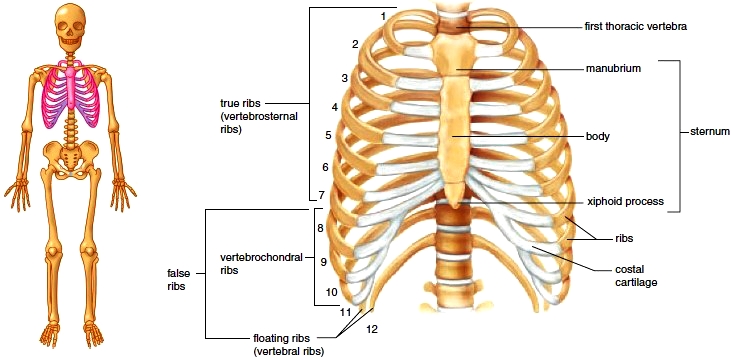
There are twelve pairs of ribs. All twelve pairs connect directly to the thoracic vertebrae in the back. After connecting with thoracic vertebrae, each rib first curves outward and then forward and downward. A rib articulates with the body of one vertebra and the transverse processes of two adjoining thoracic vertebra (called facet for tubercle of rib) (see Fig. 6.9). The upper seven pairs of ribs connect directly to the sternum by means of costal cartilages. These are called the “true ribs,” or the vertebrosternal ribs. The next three pairs of ribs are called the “false ribs,” or vertebrochondral ribs, because they attach to the sternum by means of a common cartilage. The last two pairs are called “floating ribs,” or vertebral ribs, because they do not attach to the sternum at all.
Atlas and Axis The first two cervical vertebrae are not typical (Fig. 6.9b). The atlas supports and balances the head. It has two depressions that articulate with the occipital condyles, allowing movement of the head forward and back. The axis has an odontoid process (also called the dens) that projects into the ring of the atlas. When the head moves from side to side, the atlas pivots around the odontoid process.
Sacrum and Coccyx The five sacral vertebrae are fused to form the sacrum. The sacrum articulates with the pelvic girdle and forms the posterior wall of the pelvic cavity. The coccyx, or tailbone, is the last part of the vertebral column. It is formed from a fusion of three to five vertebrae.
The Rib Cage
The rib cage (Fig. 6.10), sometimes called the thoracic cage, is composed of the thoracic vertebrae, ribs and associated cartilages, and sternum. The rib cage demonstrates how the skeleton is protective but also flexible. The rib cage protects the heart and lungs; yet it swings outward and upward upon inspiration and then downward and inward upon expiration. The rib cage also provides support for the bones of the pectoral girdle.
The Ribs
There are twelve pairs of ribs. All twelve pairs connect directly to the thoracic vertebrae in the back. After connecting with thoracic vertebrae, each rib first curves outward and then forward and downward. A rib articulates with the body of one vertebra and the transverse processes of two adjoining thoracic vertebra (called facet for tubercle of rib) (see Fig. 6.9). The upper seven pairs of ribs connect directly to the sternum by means of costal cartilages. These are called the “true ribs,” or the vertebrosternal ribs. The next three pairs of ribs are called the “false ribs,” or vertebrochondral ribs, because they attach to the sternum by means of a common cartilage. The last two pairs are called “floating ribs,” or vertebral ribs, because they do not attach to the sternum at all.
The Sternum
The sternum, or breastbone, is a flat bone that has the shape of a blade. The sternum, along with the ribs, helps protect the heart and lungs. During surgery the sternum may be split to allow access to the organs of the thoracic cavity. The sternum is composed of three bones that fuse during fetal development. These bones are the manubrium, the body, and the xiphoid process. The manubrium is the superior portion of the sternum. The body is the middle and largest part of the sternum, and the xiphoid process is the inferior and smallest portion of the sternum. The manubrium joins with the body of the sternum at an angle. This joint is an important anatomical landmark because it occurs at the level of the second rib, and therefore allows the ribs to be counted. Counting the ribs is sometimes done to determine where the apex of the heart is located-usually between the fifth and sixth ribs. The manubrium articulates with the costal cartilages of the first and second ribs; the body articulates costal cartilages of the second through tenth ribs; and the xiphoid process doesn’t articulate with any ribs. The xiphoid process is the third part of the sternum. Composed of hyaline cartilage in the child, it becomes ossified in the adult. The variably shaped xiphoid process serves as an attachment site for the diaphragm, which separates the thoracic cavity from the abdominal cavity.
The sternum, or breastbone, is a flat bone that has the shape of a blade. The sternum, along with the ribs, helps protect the heart and lungs. During surgery the sternum may be split to allow access to the organs of the thoracic cavity. The sternum is composed of three bones that fuse during fetal development. These bones are the manubrium, the body, and the xiphoid process. The manubrium is the superior portion of the sternum. The body is the middle and largest part of the sternum, and the xiphoid process is the inferior and smallest portion of the sternum. The manubrium joins with the body of the sternum at an angle. This joint is an important anatomical landmark because it occurs at the level of the second rib, and therefore allows the ribs to be counted. Counting the ribs is sometimes done to determine where the apex of the heart is located-usually between the fifth and sixth ribs. The manubrium articulates with the costal cartilages of the first and second ribs; the body articulates costal cartilages of the second through tenth ribs; and the xiphoid process doesn’t articulate with any ribs. The xiphoid process is the third part of the sternum. Composed of hyaline cartilage in the child, it becomes ossified in the adult. The variably shaped xiphoid process serves as an attachment site for the diaphragm, which separates the thoracic cavity from the abdominal cavity.

Figure 6.10 The rib cage. This structure includes the thoracic vertebrae, the ribs, and the sternum. The three bones thatmake up the sternum are the manubrium, body, and xiphoid process. The ribs numbered 1-7 are true ribs; those numbered 8-12 are false ribs.
Contacts: lubopitno_bg@abv.bg www.encyclopedia.lubopitko-bg.com Corporation. All rights reserved.
DON'T FORGET - KNOWLEDGE IS EVERYTHING!