Bones of the Appendicular Skeleton
The appendicular skeleton may be considered in two divisions: upper and lower. The upper division on each side includes the shoulder, the arm (between the shoulder and the elbow), the forearm (between the elbow and the wrist), the wrist, the hand, and the fingers. The lower division includes the hip (part of the pelvic girdle), the thigh (between the hip and the knee), the leg (between the knee and the ankle), the ankle, the foot, and the toes.
The Upper Division of the Appendicular Skeleton
The bones of the upper division may be divided into two groups, the shoulder girdle and the upper extremity.
The Shoulder Girdle The shoulder girdle consists of two bones (Fig. 3-15):
* The clavicle, or collarbone, is a slender bone with two shallow curves. It joins the sternum anteriorly and the scapula laterally and helps to support the shoulder. Because it often receives the full force of falls on outstretched arms or of blows to the shoulder, it is the most frequently broken bone.

Figure 3-15 The shoulder girdle and scapula. (A) Bones of the shoulder girdle, left anterior view. (B) Left scapula, posterior view.
* The scapula, or shoulder blade, is shown from anterior and posterior views in Figure 3-15. The spine of the scapula is the posterior raised ridge that can be felt behind the shoulder in the upper portion of the back. Muscles that move the arm attach to fossae (depressions), known as the supraspinous fossa and the infraspinous fossa, superior and inferior to the scapular spine. The acromion is the process that joins the clavicle. This can be felt as the highest point of the shoulder. Below the acromion there is a shallow socket, the glenoid cavity, that forms a ball-and-socket joint with the arm bone (humerus). Medial to the glenoid cavity is the coracoid process, to which muscles attach.
The Upper Extremity The upper extremity is also referred to as the upper limb, or simply the arm, although technically, the arm is only the region between the shoulder and the elbow. The region between the elbow and wrist is the forearm. The upper extremity consists of the following bones:
* The proximal bone is the humerus, or arm bone (Fig. 3-16). The head of the humerus forms a joint with the glenoid cavity of the scapula. The distal end has a projection on each side, the medial and lateral epicondyles, to which tendons attach, and a pulley-shaped midportion, the trochlea, that forms a joint with the ulna of the forearm.
* The proximal bone is the humerus, or arm bone (Fig. 3-16). The head of the humerus forms a joint with the glenoid cavity of the scapula. The distal end has a projection on each side, the medial and lateral epicondyles, to which tendons attach, and a pulley-shaped midportion, the trochlea, that forms a joint with the ulna of the forearm.
* The forearm bones are the ulna and the radius. In the anatomic position, the ulna lies on the medial side of the forearm in line with the little finger, and the radius lies laterally, above the thumb (Fig. 3-17). When the forearm is supine, with the palm up or forward, the two bones are parallel; when the forearm is prone, with the palm down or back, the distal end of the radius rotates around the ulna so that the shafts of the two bones are crossed (Fig. 3-18). In this position, a distal projection (styloid process) of the ulna pops up at the outside of the wrist.
Figure 3-16 The right humerus.
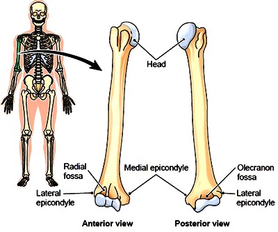

Figure 3-17 Radius and ulna of the right forearm.
* The proximal end of the ulna has the large olecranon that forms the point of the elbow (see Fig. 3-17). The trochlea of the distal humerus fits into the deep trochlear notch of the ulna, allowing a hinge action at the elbow joint. This ulnar depression, because of its deep half-moon shape, is also known as the semilunar notch (Fig. 3-19).
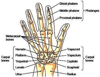
* The wrist contains eight small carpal bones arranged in two rows of four each. The names of these eight different bones are given in Figure 3-20.
* Five metacarpal bones are the framework for the palm of each hand. Their rounded distal ends form the knuckles.
* There are 14 phalanges, or finger bones, in each hand, two for the thumb and three for each finger. Each of these bones is called a phalanx.
They are identified as the first, or proximal, which is attached to a metacarpal; the second, or middle; and the third, or distal. Note that the thumb has only two phalanges, a proximal and a distal (see Fig. 3-20).
Figure 3-19 Left elbow, lateral view.

Figure 3-20 Bones of the right hand, anterior view.
Figure 3-18 Movements of the forearm. When the palm is supine (facing up or forward), the radius and ulna are parallel. When the palm is prone (facing down or to the rear), the radius crosses over the ulna.

Contacts: lubopitno_bg@abv.bg www.encyclopedia.lubopitko-bg.com Corporation. All rights reserved.
DON'T FORGET - KNOWLEDGE IS EVERYTHING!