Skeletal Muscles of the Body
The human body has some 600 skeletal muscles, but this text will discuss only some of the most significant of these. First, let us consider certain basic principles of muscle contraction.
Basic Principles
When a muscle contracts, one bone remains fairly stationary, and the other one moves. The origin of a muscle is on the stationary bone, and the insertion of a muscle is on the bone that moves. Frequently, a body part is moved by a group of muscles working together. Even so, one muscle does most of the work, and this muscle is called the prime mover. For example, in flexing the elbow, the prime mover is the biceps brachii (Fig. 7.10) The assisting muscles are called the synergists. The brachialis (see Fig. 7.12) is a synergist that helps the biceps brachii flex the elbow. A prime mover can have several synergists. When muscles contract, they shorten. Therefore, muscles can only pull; they cannot push. However, muscles have antagonists, and antagonistic pairs work opposite one another to bring about movement in opposite directions. For example, the biceps brachii and the triceps brachii are antagonists; one flexes the forearm, and the other extends the forearm (Fig. 7.10). Later on in our discussion, we will encounter other antagonistic pairs.
Figure 7.10 The origin of a muscle is on a bone that remains stationary, and the insertion of a muscle is on a bone that moves when a muscle contracts. Two of the muscles shown here are antagonistic. a. When the biceps brachii contracts, the lower arm flexes. b. When the triceps brachii contracts, the lower arm extends.
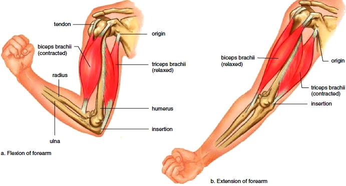
Figure 7.12 Posterior view of the body’s superficial skeletal muscles.
Naming Muscles
When learning the names of muscles, considering what the name means will help you remember it. The names of the various skeletal muscles are often combinations of the following terms used to characterize muscles:
1. Size. For example, the gluteus maximus is the largest muscle that makes up the buttocks. The gluteus minimus is the smallest of the gluteal muscles. Other terms used to indicate size are vastus (huge), longus (long), and brevis (short).
2. Shape. For example, the deltoid is shaped like a delta, or triangle, while the trapezius is shaped like a trapezoid. Other terms used to indicate shape are latissimus (wide) and teres (round).
3. Direction of fibers. For example, the rectus abdominis is a longitudinal muscle of the abdomen (rectus means straight). The orbicularis is a circular muscle around the eye. Other terms used to indicate direction are transverse (across) and oblique (diagonal).
4. Location. For example, the frontalis overlies the frontal bone. The external obliques are located outside the internal obliques. Other terms used to indicate location are pectoralis (chest), gluteus (buttock), brachii (arm), and sub (beneath). You should also review these directional terms: anterior, posterior, lateral, medial, proximal, distal, superficial, and deep.
5. Attachment. For example, the sternocleidomastoid is attached to the sternum, clavicle, and mastoid process. The brachioradialis is attached to the brachium (arm) and the radius.
6. Number of attachments. For example, the biceps brachii has two attachments, or origins (and is located on the arm). The quadriceps femoris has four origins (and is located on the anterior femur).
7. Action. For example, the extensor digitorum extends the fingers or digits. The adductor magnus is a large muscle that adducts the thigh. Other terms used to indicate action are flexor (to flex), masseter (to chew), and levator (to lift).
Figure 7.11 Anterior view of the body’s superficial skeletal muscles.
Skeletal Muscle Groups
In our discussion, the muscles of the body (Figs. 7.11 and 7.12) will be grouped according to their location and their action. After you understand the meaning of a muscle’s name, try to correlate its name with the muscle’s location and the action it performs. Knowing the origin and insertion will also
help you remember what the muscle does. Why? Because the insertion is on the bone that moves. Only then will you be able to understand the actions of the muscles listed in Tables 7.2. Scientific terminology is necessary because it allows all persons to know the exact action being described for that muscle. Also review the meaning of the terms arm and leg.
Muscles of the Head
The muscles of the head and neck are the first group of muscles we will study. The muscles of the head and neck are illustrated in Figure 7.13 and listed in Table 7.2. The muscles of the head are responsible for facial expression and mastication (chewing). One muscle of the head and several muscles of the neck allow us to swallow. The muscles of the neck also move the head.
Muscles of Facial Expression
The muscles of facial expression are located on the scalp and face. These muscles are unusual in that they insert into and move the skin. Therefore, we expect them to move the skin and not a bone. The use of these muscles communicates to others whether we are surprised, angry, fearful, happy, and so forth.
Frontalis lies over the frontal bone; it raises the eyebrows and wrinkles the brow. Frequent use results in furrowing of the forehead.
Orbicularis oculi is a ringlike band of muscle that encircles (forms an orbit about) the eye. It causes the eye to close or blink, and is responsible for “crow’s feet” at the eye corners.
Orbicularis oris encircles the mouth and is used to pucker the lips, as in forming a kiss. Frequent use results in lines about the mouth.
Buccinator muscles are located in the cheek areas. When a buccinator contracts, the cheek is compressed, as when a person whistles or blows out air. Therefore, this muscle is called the “trumpeter’s muscle.” Important to everyday life, the buccinator helps hold food in contact with the teeth during chewing. It is also used in swallowing, as discussed next.
Zygomaticus extends from each zygomatic arch (cheekbone) to the corners of the mouth. It raises the corners of the mouth when a person smiles.
Muscles of Mastication
The muscles of mastication are used when we chew food or bite something. Although there are four pairs of muscles for chewing, only two pairs are superficial and shown in Figure 7.13. As you might expect, both of these muscles insert on the mandible. Each masseter has its origin on the zygomatic arch and its insertion on the mandible. The masseter is a muscle of mastication (chewing) because it is a prime mover for elevating the mandible. Each temporalis is a fan-shaped muscle that overlies the temporal bone. It is also a prime mover for elevating the mandible. The masseter and temporalis are synergists.
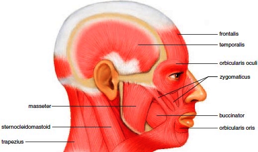
Figure 7.13 Muscles of the head and neck. Some of these muscles account for our facial expressions and the ability to chew our food; others move the head.


Contacts: lubopitno_bg@abv.bg www.encyclopedia.lubopitko-bg.com Corporation. All rights reserved.
DON'T FORGET - KNOWLEDGE IS EVERYTHING!