Figure 3.10 The cell cycle consists of interphase, during which cellular components duplicate, and a mitotic stage, during which the cell divides. Interphase consists of two so-called “growth” phases (G1 and G2) and a DNA synthesis (S) phase. The mitotic stage consists of the phases noted plus cytokinesis.
The Cell Cycle
The cell cycle is an orderly set of stages that take place between the time a cell divides and the time the resulting daughter cells also divide. The cell cycle is controlled by internal and external signals. A signal is a molecule that stimulates or inhibits a metabolic event. For example, growth factors are external signals received at the plasma membrane that cause a resting cell to undergo the cell cycle. When blood platelets release a growth factor, skin fibroblasts in the vicinity finish the cell cycle, thereby repairing an injury. Other signals ensure that the stages follow one another in the normal sequence and that each stage of the cell cycle is properly completed before the next stage begins. The cell cycle has a number of checkpoints, places where the cell cycle stops if all is not well.
Any cell that did not successfully complete mitosis and is abnormal undergoes apoptosis at the restriction checkpoint. Apoptosis is often defined as programmed cell death because the cell progresses through a series of events that bring about its destruction. The cell rounds up and loses contact with its neighbors. The nucleus fragments, and the plasma membrane develops blisters. Finally, the cell fragments, and its bits and pieces are engulfed by white blood cells and/or neighboring cells. The enzymes that bring about apoptosis are ordinarily held in check by inhibitors, but are unleashed by either internal or external signals. Following a certain number of cell cycle revolutions, cells are apt to become specialized and no longer go through the cell cycle. Muscle cells and nerve cells typify specialized cells that rarely, if ever, go through the cell cycle. At the other extreme, some cells in the body, called stem cells, are always immature and go through the cell cycle repeatedly. There is a great deal of interest in stem cells today because it may be possible to control their future development into particular tissues and organs.
The cell cycle has two major portions: interphase and the mitotic stage (Fig. 3.10).
Interphase
The cell in Figure 3.1 is in interphase because it is not dividing. During interphase, the cell carries on its regular activities, and it also gets ready to divide if it is going to complete the cell cycle. For these cells, interphase has three stages, called G1 phase, S phase, and G2 phase. G1 Phase Early microscopists named the phase before DNA replication G1, and they named the phase after DNA replication G2. G stood for “gap.” Now that we know how metabolically active the cell is, it is better to think of G as standing for “growth.” Protein synthesis is very much a part of these growth phases. During G1, a cell doubles its organelles (such as mitochondria and ribosomes) and accumulates materials that will be used for DNA synthesis. S Phase Following G1, the cell enters the S (for “synthesis”) phase. During the S phase, DNA replication occurs. At the beginning of the S phase, each chromosome is composed of one DNA double helix, which is equal to a chromatid. At the end of this phase, each chromosome has two identical DNA double helix molecules, and therefore is composed of two sister chromatids. Another way of expressing these events is to say that DNA replication has resulted in duplicated chromosomes. G2 Phase During this phase, the cell synthesizes proteins that will assist cell division, such as the protein found in microtubules. Also, chromatin condenses, and the chromosomes become visible.
Mitotic Stage
Following interphase, the cell enters the M (for mitotic) stage. This cell division stage includes mitosis (division of the nucleus) and cytokinesis (division of the cytoplasm). During mitosis, daughter chromosomes are distributed to two daughter nuclei. When cytokinesis is complete, two daughter cells are present.
Events During Interphase
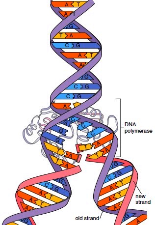
Two significant events during interphase are replication of DNA and protein synthesis. Replication of DNA During replication, an exact copy of a DNA helix is produced. The double-stranded structure of DNA aids replication because each strand serves as a template for the formation of a complementary strand. A template is most often a mold used to produce a shape opposite to itself. In this case, each old (parental) strand is a template for each new (daughter) strand. Figures 3.11 and 3.12 show how replication is carried out. Figure 3.12 uses the ladder configuration of DNA for easy viewing.
1. Before replication begins, the two strands that make up parental DNA are hydrogen-bonded to one another.
2. During replication, the old (parental) DNA strands unwind and “upzip” (i.e., the weak hydrogen bonds between the two strands break).
3. New complementary nucleotides, always present in the nucleus, pair with the nucleotides in the old strands. A pairs with T and C pairs with G. The enzyme DNA polymerase joins the new nucleotides forming new (daughter) complementary strands.
4. When replication is complete, the two double helix molecules are identical.
Each strand of a double helix is equal to a chromatid, which means that at the completion of replication each chromosome is composed of two sister chromatids. They are called sister chromatids because they are identical. The chromosome is called a duplicated chromosome.
Cancer, which is characterized by rapidly dividing cells, is treated with chemotherapeutic drugs that stop replication and therefore cell division. Some chemotherapeutic drugs are analogs that have a similar, but not identical, structure to the four nucleotides in DNA. When these are mistakenly used by the cancer cells to synthesize DNA, replication stops, and the cells die off.
Cell Cycle Stages
The cell cycle has two major portions: interphase and the mitotic stage (Fig. 3.10).
Interphase
The cell in Figure 3.1 is in interphase because it is not dividing. During interphase, the cell carries on its regular activities, and it also gets ready to divide if it is going to complete the cell cycle. For these cells, interphase has three stages, called G1 phase, S phase, and G2 phase. G1 Phase Early microscopists named the phase before DNA replication G1, and they named the phase after DNA replication G2. G stood for “gap.” Now that we know how metabolically active the cell is, it is better to think of G as standing for “growth.” Protein synthesis is very much a part of these growth phases. During G1, a cell doubles its organelles (such as mitochondria and ribosomes) and accumulates materials that will be used for DNA synthesis. S Phase Following G1, the cell enters the S (for “synthesis”) phase. During the S phase, DNA replication occurs. At the beginning of the S phase, each chromosome is composed of one DNA double helix, which is equal to a chromatid. At the end of this phase, each chromosome has two identical DNA double helix molecules, and therefore is composed of two sister chromatids. Another way of expressing these events is to say that DNA replication has resulted in duplicated chromosomes. G2 Phase During this phase, the cell synthesizes proteins that will assist cell division, such as the protein found in microtubules. Also, chromatin condenses, and the chromosomes become visible.
Mitotic Stage
Following interphase, the cell enters the M (for mitotic) stage. This cell division stage includes mitosis (division of the nucleus) and cytokinesis (division of the cytoplasm). During mitosis, daughter chromosomes are distributed to two daughter nuclei. When cytokinesis is complete, two daughter cells are present.
Events During Interphase
Two significant events during interphase are replication of DNA and protein synthesis. Replication of DNA During replication, an exact copy of a DNA helix is produced. The double-stranded structure of DNA aids replication because each strand serves as a template for the formation of a complementary strand. A template is most often a mold used to produce a shape opposite to itself. In this case, each old (parental) strand is a template for each new (daughter) strand. Figures 3.11 and 3.12 show how replication is carried out. Figure 3.12 uses the ladder configuration of DNA for easy viewing.
1. Before replication begins, the two strands that make up parental DNA are hydrogen-bonded to one another.
2. During replication, the old (parental) DNA strands unwind and “upzip” (i.e., the weak hydrogen bonds between the two strands break).
3. New complementary nucleotides, always present in the nucleus, pair with the nucleotides in the old strands. A pairs with T and C pairs with G. The enzyme DNA polymerase joins the new nucleotides forming new (daughter) complementary strands.
4. When replication is complete, the two double helix molecules are identical.
Each strand of a double helix is equal to a chromatid, which means that at the completion of replication each chromosome is composed of two sister chromatids. They are called sister chromatids because they are identical. The chromosome is called a duplicated chromosome.
Cancer, which is characterized by rapidly dividing cells, is treated with chemotherapeutic drugs that stop replication and therefore cell division. Some chemotherapeutic drugs are analogs that have a similar, but not identical, structure to the four nucleotides in DNA. When these are mistakenly used by the cancer cells to synthesize DNA, replication stops, and the cells die off.
Figure 3.11 Overview of DNA replication. During replication, an old strand serves as a template for a new strand. The new double helix is composed of an old (parental) strand and a new (daughter) strand.


Figure 3.12 Ladder configuration and DNA replication. Use of the ladder configuration better illustrates how complementary nucleotides available in the cell pair with those of each old strand before they are joined together to form a daughter strand.
Contacts: lubopitno_bg@abv.bg www.encyclopedia.lubopitko-bg.com Corporation. All rights reserved.
DON'T FORGET - KNOWLEDGE IS EVERYTHING!