Joints (Articulations)
Bones articulate at the joints, which are often classified according to the amount of movement they allow:
Fibrous joints are immovable. Fibrous connective tissue joins bone to bone.
Cartilaginous joints are slightly movable. Fibrocartilage is located between two bones.
Synovial joints are freely movable. In these joints, the bones do not come in contact with each other.
Fibrous Joints
Some bones, such as those that make up the cranium, are sutured together by a thin layer of fibrous connective tissue and are immovable. Review Figures 6.6 and 6.7, and note the following immovable sutures: coronal suture, between the parietal bones and the frontal bone; lambdoidal suture, between the parietal bones and the occipital bone; squamosal suture, between each parietal bone and each temporal bone; sagittal suture, between the parietal bones.
Cartilaginous Joints
Slightly movable joints are those in which the bones are joined by fibrocartilage. The ribs are joined to the sternum by costal cartilages (see Fig. 6.10). The bodies of adjacent vertebrae are separated by intervertebral disks (see Fig. 6.8) that increase vertebral flexibility. The pubic symphysis, which occurs between the pubic bones (see Fig. 6.15), consists largely of fibrocartilage. Due to hormonal changes, this joint becomes more flexible during late pregnancy, which allows the pelvis to expand during childbirth.
Synovial Joints
All synovial joints are freely movable because, unlike the joints discussed so far, the two bones are separated by a joint cavity (Figs. 6.19 and 6.20). The cavity is lined by a synovial membrane, which produces synovial fluid, a lubricant for the joint. The absence of tissue between the articulating bones allows them to be freely movable but means that the joint has to be stabilized in some way. The joint is stabilized by the joint capsule, a sleevelike extension of the periosteum of each articulating bone. Ligaments, which are composed of dense regular connective tissue, bind the two bones to one another and add even more stability. Tendons, which are cords of dense fibrous connective tissue that connect muscle to bone, also help stabilize a synovial joint. The articulating surfaces of the bones are protected in several ways. The bones are covered by a layer of articular (hyaline) cartilage. In addition, the joint, such as the knee, contains menisci (sing., meniscus), crescent-shaped pieces of cartilage and fluid-filled sacs called bursae, which ease friction between all parts of the joint. Inflammation of the bursae is called bursitis. Tennis elbow is a form of bursitis.
All synovial joints are freely movable because, unlike the joints discussed so far, the two bones are separated by a joint cavity (Figs. 6.19 and 6.20). The cavity is lined by a synovial membrane, which produces synovial fluid, a lubricant for the joint. The absence of tissue between the articulating bones allows them to be freely movable but means that the joint has to be stabilized in some way. The joint is stabilized by the joint capsule, a sleevelike extension of the periosteum of each articulating bone. Ligaments, which are composed of dense regular connective tissue, bind the two bones to one another and add even more stability. Tendons, which are cords of dense fibrous connective tissue that connect muscle to bone, also help stabilize a synovial joint. The articulating surfaces of the bones are protected in several ways. The bones are covered by a layer of articular (hyaline) cartilage. In addition, the joint, such as the knee, contains menisci (sing., meniscus), crescent-shaped pieces of cartilage and fluid-filled sacs called bursae, which ease friction between all parts of the joint. Inflammation of the bursae is called bursitis. Tennis elbow is a form of bursitis.
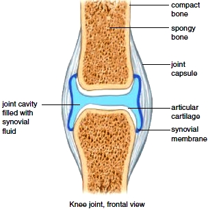
Figure 6.19 Generalized anatomy of a synovial joint.

Figure 6.20 The knee joint. Notice the menisci and bursae associated with the knee joint.
Types of Synovial Joints
Different types of freely movable joints are listed here and depicted in Figure 6.21.
Saddle joint. Each bone is saddle-shaped and fits into the complementary regions of the other. A variety of movements are possible. Example: the joint between the carpal and metacarpal bones of the thumb.
Ball-and-socket joint. The ball-shaped head of one bone fits into the cup-shaped socket of another. Movement in all planes, as well as rotation, are possible. Examples: the shoulder and hip joints.
Pivot joint. A small, cylindrical projection of one bone pivots within the ring formed of bone and ligament of another bone. Only rotation is possible. Examples: the joint between the proximal ends of the radius and ulna, and the joint between the atlas and axis.
Hinge joint. The convex surface of one bone articulates with the concave surface of another. Up-and-down motion in one plane is possible. Examples: the elbow and knee joints.
Gliding joint. Flat or slightly curved surfaces of bones articulate. Sliding or twisting in various planes is possible. Examples: the joints between the bones of the wrist and between the bones of the ankle.
Condyloid joint. The oval-shaped condyle of one bone fits into the elliptical cavity of another. Movement in different planes is possible, but rotation is not. Examples: the joints between the metacarpals and phalanges.
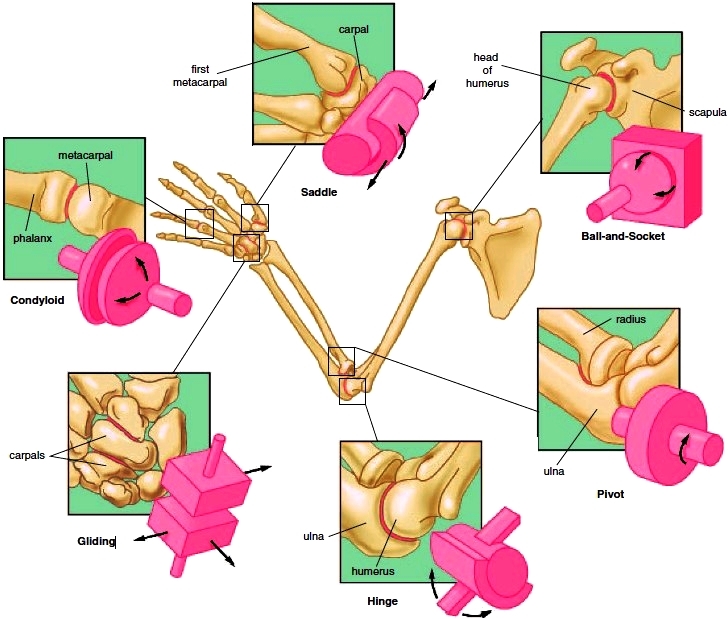
Figure 6.21 Types of synovial joints.
Movements Permitted by Synovial Joints
Skeletal muscles are attached to bones by tendons that cross joints. When a muscle contracts, one bone moves in relation to another bone. The more common types of movements are described here.
Angular Movements (Fig. 6.22a):
Flexion decreases the joint angle. Flexion of the elbow moves the forearm toward the arm; flexion of the knee moves the leg toward the thigh. Dorsiflexion is flexion of the foot upward, as when you stand on your heels; plantar flexion is flexion of the foot downward, as when you stand on your toes.
Extension increases the joint angle. Extension of the flexed elbow straightens the upper limb. Hyperextension occurs when a portion of the body part is extended beyond 180°. It is possible to hyperextend the head and the trunk of the body, and also the shoulder and wrist (arm and hand).
Adduction is the movement of a body part toward the midline. For example, adduction of the arms or legs moves them back to the sides, toward the body.
Abduction is the movement of a body part laterally, away from the midline. Abduction of the arms or legs moves them laterally, away from the body.
Circular Movements (Fig. 6.22b):
Circumduction is the movement of a body part in a wide circle, as when a person makes arm circles. Careful observation of the motion reveals that, because the proximal end of the arm is stationary, the shape outlined by the arm is actually a cone.
Rotation is the movement of a body part around its own axis, as when the head is turned to answer “no” or when the arm is twisted toward the trunk (medial rotation) and away from the trunk (lateral rotation).
Supination is the rotation of the forearm so that the palm is upward; pronation is the opposite-the movement of the forearm so that the palm is downward.
Special movements (Fig. 6.22c):
Inversion and eversion apply only to the feet. Inversion is turning the foot so that the sole faces inward, and eversion is turning the foot so that the sole faces outward.
Elevation and depression refer to the lifting up and down, respectively, of a body part, as when you shrug your shoulders or move your jaw up and down.
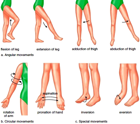
Figure 6.22 Joint movements. a. Angular movements increase or decrease the angle between the bones of a joint. b. Circular movements describe a circle or part of a circle. c. Special movements are unique to certain joints.
Contacts: lubopitno_bg@abv.bg www.encyclopedia.lubopitko-bg.com Corporation. All rights reserved.
DON'T FORGET - KNOWLEDGE IS EVERYTHING!